Comparative Study
. 2005 May 31;102(22):7934-9.
doi: 10.1073/pnas.0500357102. Epub 2005 May 20.

A critical role of T cell antigen receptor-transduced MHC class I-restricted helper T cells in tumor protection

Affiliations
Comparative Study

A critical role of T cell antigen receptor-transduced MHC class I-restricted helper T cells in tumor protection

Emma C Morris et al. Proc Natl Acad Sci U S A. .

Abstract

Adoptive transfer of antigen-specific CD4(+) and CD8(+) T cells is one of the most efficient forms of cancer immunotherapy. However, the isolation of antigen-specific CD4(+) T cells is limited because only few tumor-associated helper epitopes are identified. Here, we used T cell antigen receptor gene transfer to target CD4(+) T cells against an MHC class I-presented epitope of a model tumor antigen. IFN-gamma-producing CD4(+) T cells were unable to expand in vivo and to provide help for tumor rejection. In contrast, CD4(+) T cells producing high levels of IL-2 expanded in vivo, provided help for cytotoxic T lymphocyte-mediated tumor rejection, and developed T cell memory. The data demonstrate in vivo synergy between T cell antigen receptor-transduced CD4(+) and CD8(+) T cells specific for the same epitope resulting in long-term tumor protection.


Figures

Fig. 1.
Fig. 1.
Retroviral gene transfer of TCR chains, purification of TCR-expressing CD4+ and CD8+ T cells, and retroviral gene transfer of CD8α.(AF) Flow cytometric analysis of viable CD3+ murine splenocytes 2 days after retroviral transfer with F5 TCRα and TCRβ genes (AC) or mock transduction (DF). Cells were stained with PE-anti-Vβ11, PE-labeled NP tetramer, FITC-anti-CD4, or allophycocyanin-anti-CD8. (G and H) Flow cytometric analysis of purified CD4+ and CD8+ T cells. Forty-eight hours after transduction, TCR-td bulk T cells were sorted into CD4+ and CD8+ T cells by using magnetic beads, followed by enrichment for Vβ11+ cells 1 day later. Cells were stained with PE-anti-Vβ11, FITC-anti-CD4, and/or allophycocyanin-anti-CD8, and quadrants were set based on anti-CD4 and anti-CD8 stained mock-td cells. (I and J) Flow cytometric analysis of purified TCR-td CD4+ T cells (I) and TCR-td CD4+8+ T cells (J) after retroviral cotransfer of F5 TCRα, TCRβ, and murine CD8α genes. After cotransfer of CD8α, >95% of CD4+ T cells coexpressed CD8α (TCR-td CD4+8+ T cells).
Fig. 2.
Fig. 2.
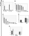
In vitro functional analysis of TCR-td bulk T cells, CD8+ T cells, CD4+ T cells, and CD4+8+ T cells. (A) Peptide-specific IFN-γ secretion of TCR-td bulk T cells (black bars) in response to peptide-loaded DCs and MHC class-II negative peptide-loaded EL4 tumor cells. Mock-td bulk T cells (gray bars) did not secrete IFN-γ in response to any of the targets. (B) Purified populations of TCR-td CD8+ T cells (black bars) and TCR-td CD4+ T cells (gray bars) secreted IFN-γ in response to peptide-loaded syngeneic DCs. TCR-td CD8+ T cells recognized 10 pM peptide, whereas a 10-fold higher concentration of peptide (100 pM) was required to trigger IFN-γ secretion by the TCR-td CD4+ T cells. (C) Using peptide-loaded EL4 tumor cells as targets, TCR-td CD8+ T cells (black bars) mounted an efficient IFN-γ response, whereas the TCR-td CD4+ T cells (gray bars) did not. (D) Transcostimulation by means of the addition of syngeneic DCs rescued the peptide-specific IFN-γ response of TCR-td CD4+ T cells (gray bars) to MHC class II negative NP-expressing EL4 tumor cells (EL4 NP) compared with that observed with TCR-td CD8+ T cells (black bars). (E) Coexpression of murine CD8α abolished the DC dependence of the IFN-γ response. TCR-td CD4+8+ T cells (gray bars) and TCR-td CD8+ T cells (black bars) respond to antigen presented by DCs and EL4 NP.
Fig. 3.
Fig. 3.
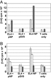
In vitro proliferative potential and IL-2 production of TCR-td CD4+ T cells and TCR-td CD4+8+ T cells. (A) TCR-td CD4+8+ T cells proliferated poorly in response to peptide-presenting tumor cells (white bars) compared with TCR-td CD4+ T cells (gray bars) and TCR-td CD8+ T cells (black bars). The proliferative potential of the TCR-td T cell populations correlated well with IL-2 production. (B) TCR-td CD4+8+ T cells (white bars) secreted reduced levels of IL-2 in response to EL4 NP tumor cells as compared with TCR-td CD4+ T cells (gray bars). TCR-td CD8+ T cells (black bars) did not secrete IL-2 in response to any of the targets. Proliferation of the IL-2-dependent cell line CTLL was used to measure the IL-2 content in the supernatant taken from the stimulated T cells.
Fig. 4.
Fig. 4.
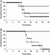
TCR-td CD4+ T cells provide in vivo help for CD8 T cell-mediated tumor rejection. C57BL/6 mice received nonmyeloablative radiation (600 rad) and then 12 h later were injected s.c. with 3 × 10⁶ EL4 NP tumor cells. (A) A further 24 h later, mice received 106 TCR-td CD4+ T cells with 104 TCR-td CD8+ T cells (open triangles), 106 TCR-td CD4+8+ T cells with 104 TCR-td CD8+ T cells (crosses), 106 mock-td bulk T cells (open squares), or PBS (diamonds). (B) In a separate tumor protection experiment, mice were conditioned and tumor-challenged as above, followed by transfer of 106 mock-td CD4+ T cells with 104 TCR-td CD8+ T cells (open squares), 106 TCR-td CD4+ T cells with anti-CD8-blocking antibody (filled diamonds), or 106 TCR-td CD4+ T cells with 2 × 10⁴ TCR-td CD8+ T cells (open triangles).
Fig. 5.
Fig. 5.
Detection and in vivo persistence of TCR-td T cells in tumor-bearing mice. Shown is the analysis of splenocytes prepared from mice in Fig. 4A at the time they were killed because of tumor burden. Adoptively transferred T cells were identified by triple staining with antibodies specific for Thy1.1, Vβ11, and CD4. Representative examples are shown for each group of mice, and all FACS plots display viable lymphocytes gated on Vβ11+ T cells. (A) No Thy1.1+ Vβ11+ T cells were identified in four tumor-bearing PBS-treated control mice. (B) In four mice treated with mock-td CD4+ T cells, between 0.5% and 1.7% of the gated Vβ11+ cells were the CD4+ Thy1.1+ transferred T cells. (C) Similar numbers of TCR-td CD4+8+ cells were detected in three mice (0.5–3.3%) suggesting a lack of antigen-driven expansion of the TCR-td CD4+8+ as compared with the Mock-td CD4+ T cells. (D) Analysis of the spleen of a tumor-bearing animal that received TCR-td CD4+ T cells showed a large expansion of transferred Thy1.1+ CD4+ T cells (34.0% of gated Vβ11+), together with an expansion of the cotransferred Thy1.1+ CD8+ T cells (13.6% of gated Vβ11+). Staining of lymph node cells of mice in the four treatment groups showed similar levels of transferred Thy1.1+ Vβ11+ CD4+ T cells (data not shown).
Fig. 6.
Fig. 6.
TCR-td CD4+ T cells persist and develop long-term memory in vivo. Shown is the analysis of splenocytes and lymph node cells prepared from tumor-free mice in Fig. 4A on day 35 or day 95 after T cell transfer. Adoptively transferred T cells were identified by triple staining with antibodies specific for Thy1.1, Vβ11, and CD4, and all FACS plots display viable lymphocytes gated on Vβ11+ T cells. (A and B) A day-35 comparison of splenocytes of mice treated with TCR-td CD4+ (A) and CD4+8+ (B) cells. Similar frequencies of transferred Thy1.1+ CD4+ T cells were detected in lymph node samples (data not shown). (CH) At day 90, three tumor-free mice were challenged s.c. with 1 × 10⁶ irradiated EL4 NP tumor cells in the right lower leg. After 5 days, ex vivo FACS analysis was used to analyze T cell responses in the tumor-antigen-exposed right inguinal and popliteal lymph nodes, by using the nonexposed left inguinal and popliteal lymph nodes as a baseline control. The TCR-td CD4+8+ T cells did not respond to tumor rechallenge at day 90 (C and D), whereas both mice that received TCR-td CD4+ cells demonstrated an expansion of TCR-td CD4+ and CD8+ T cells in the lymph nodes of the tumor antigen exposed right side (EH).

Similar articles

Cited by

References

    1. Rooney, C. M., Smith, C. A., Ng, C. Y., Loftin, S., Li, C., Krance, R. A., Brenner, M. K. & Heslop, H. E. (1995) Lancet 345, 9–13. - PubMed
    1. Rooney, C. M., Smith, C. A., Ng, C. Y., Loftin, S. K., Sixbey, J. W., Gan, Y., Srivastava, D. K., Bowman, L. C., Krance, R. A., Brenner, M. K. & Heslop, H. E. (1998) Blood 92, 1549–1555. - PubMed
    1. Khanna, R., Bell, S., Sherritt, M., Galbraith, A., Burrows, S. R., Rafter, L., Clarke, B., Slaughter, R., Falk, M. C., Douglass, J., et al. (1999) Proc. Natl. Acad. Sci. USA 96, 10391–10396. - PMC - PubMed
    1. Haque, T., Taylor, C., Wilkie, G. M., Murad, P., Amlot, P. L., Beath, S., McKiernan, P. J. & Crawford, D. H. (2001) Transplantation 72, 1399–1402. - PubMed
    1. Haque, T., Wilkie, G. M., Taylor, C., Amlot, P. L., Murad, P., Iley, A., Dombagoda, D., Britton, K. M., Swerdlow, A. J. & Crawford, D. H. (2002) Lancet 360, 436–442. - PubMed

Publication types

MeSH terms