Comparative Study
. 2005 Aug;89(2):1020-9.
doi: 10.1529/biophysj.105.062240. Epub 2005 May 20.

KvAP-based model of the pore region of shaker potassium channel is consistent with cadmium- and ligand-binding experiments

Affiliations
Comparative Study

KvAP-based model of the pore region of shaker potassium channel is consistent with cadmium- and ligand-binding experiments

Iva Bruhova et al. Biophys J. 2005 Aug.

Abstract

Potassium channels play fundamental roles in excitable cells. X-ray structures of bacterial potassium channels show that the pore-lining inner helices obstruct the cytoplasmic entrance to the closed channel KcsA, but diverge in widely open channels MthK and KvAP, suggesting a gating-hinge role for a conserved Gly in the inner helix. A different location of the gating hinge and a narrower open pore were proposed for voltage-gated Shaker potassium channels that have the Pro-473-Val-Pro motif. Two major observations back the proposal: cadmium ions lock mutant Val-476-Cys in the open state by bridging Cys-476 and His-486 in adjacent helices, and cadmium blocks the locked-open double mutant Val-474-Cys/Val-476-Cys by binding to Cys-474 residues. Here we used molecular modeling to show that the open Shaker should be as wide as KvAP to accommodate an open-channel blocker, correolide. We further built KvAP-, MthK-, and KcsA-based models of the Shaker mutants and Monte-Carlo-minimized them with constraints Cys-476-Cd(2+)-His-486. The latter were consistent with the KvAP-based model, causing a small-bend N-terminal to the Pro-473-Val-Pro motif. The constraints significantly distorted the MthK-based structure, making it similar to KvAP. The KcsA structure resisted the constraints. Two Cd(2+) ions easily block the locked-open KvAP-based model at Cys-474 residues, whereas constraining a single cadmium ion to four Cys-474 caused large conformational changes and electrostatic imbalance. Although mutual disposition of the voltage-sensor and pore domains in the KvAP x-ray structure is currently disputed, our results suggest that the pore-region domain retains a nativelike conformation in the crystal.


Figures

FIGURE 1
FIGURE 1
Structure of correolide.
FIGURE 2
FIGURE 2
Dimensions of Shaker channel intracellular blockers. (A) Plots of MC-minimized energy of correolide pulled via the variable-diameter rings of methane molecules. The zero position corresponds to the epoxy group of correolide in the plane of the ring. The energy barrier increases sharply as the diameter decreases from 10 to 9.5 Å. (B) The superposition of MC-minimized structures of correolide pulled through the rings with inner diameter di of 8 and 10 Å. Large deformations of correolide in the smaller ring explain the high energy barrier shown in A. MCM caused some methane molecules of the smaller ring to violate in-plane constraints to avoid strong repulsions with their neighbors. (C) Plots of MC-minimized energy of tetrabutylammonium pulled via the variable-diameter rings of methane molecules. The zero position corresponds to the nitrogen atom in the plane of the ring. The barrier increases sharply as the diameter decreases from 6.5 to 6 Å. (D) The superposition of MC-minimized structures of tetrabutylammonium pulled through the rings inner diameter di of 6 and 8 Å.
FIGURE 3
FIGURE 3
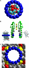
The 10-Å ring of methane molecules versus correolide and KvAP-based model of the Shaker channel. (A) Space-filled model of correolide viewed via the ring. (B) The side view of the Shaker model. The inner helices in two opposite domains are shown as ribbons. C-terminal parts of the pore helices and the selectivity-filter region are shown as rods. Correolide-sensing residues Ala-463, Val-467, Ala-471, Val-474, and Pro-475 are space-filled. Blue-colored Pro-475 at the cytoplasmic entrance is aligned with the 10-Å ring embracing correolide molecules in positions 0 through 8 Å from the ring plane (left). In the latter position (right), correolide would occur between the selectivity filter and the cytoplasmic entrance to the pore, which should be at least 10 Å wide to enable high-affinity binding of the drug. (C) Space-filled model of the KvAP-based model of the Shaker channel viewed via the 10-Å ring. Pro-473, Val-474, and Pro-475 are colored red, yellow, and green, respectively. Hydrogen atoms are not shown. Note a perfect match in the dimensions of the ring and the cytoplasmic entrance to the pore.
FIGURE 4
FIGURE 4
Convergence of the Shaker models from the x-ray-based starting structures to conformations with coordinating bonds Cys-476_Sγ—Cd2+—Nɛ2_His-486. Cd2+ ions were constrained to Sγ_Cys-476 at the distance of 2.65 Å. Distance constraints Cd2+—Nɛ2_His-486 were decreased with a step 0.5 Å from the starting values found in the x-ray-based structures. At each step, the energy was MC-minimized until the distances of ∼2.3 Å were achieved. In the plots of MC-minimized energy against the imposed distance Cd2+—Nɛ2_His-486, the energy is shown relative to the lowest-energy structure found. The locked-open conformations are more preferable energetically than the starting KvAP- and MthK-based structures. KcsA-based conformation with coordinating bonds Cys-476_Sγ—Cd2+—Nɛ2_His-486 has higher energy than the starting structure. Superposed MC-minimized structures are at the right of the respective energy plots. In the side and cytoplasmic views, only the inner helices are shown for clarity. Cys and His are shown as sticks and Cd2+ ions as magenta spheres. Locking open the KvAP-based structure is energetically preferable and requires minimal structural deformations.
FIGURE 5
FIGURE 5
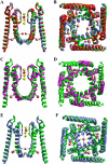
KvAP-based models of the Shaker channel. (A and B) Side and cytoplasmic views of the superposition of the x-ray based structure (red), locked-open mutant Val-476-Cys (green), and locked-open mutant Val-474-Cys/Val-476-Cys (violet) blocked by two Cd2+ ions. The side chains of Glu-395, Cys-476, and His-486 are shown as sticks, Cd2+ ions as magenta spheres, and K+ ions as yellow spheres. The involvement of Glu-395 in the Cd2+ coordination sphere was an unexpected result of MCM. The coordination occurred despite Glu-395 was not ionized in the model. In the cell, negative charges at Glu-395 and Cys-474 would facilitate Cd2+ binding. (C and D) The superposition of the locked-open conformations obtained from KvAP (green) and MthK (magenta) starting structures. The conformations are similar despite starting structures being essentially different, especially at the C-termini (Table 2). For clarity, only two opposed domains are shown in the side views and only inner and outer helices are shown in cytoplasmic views. (E and F) The superposition of the locked-open model (green) and the locked-open model blocked by a single Cd2+ ion (violet). Constraining a single Cd2+ ion to four Cys-474 residues caused large conformational deformations, which are inconsistent with the experimentally observed easiness of the Cd2+ block of the locked-open channel.
FIGURE 6
FIGURE 6
KvAP-based model of the Shaker inner-helices bundle obtained by MCM without using the advantage of the channel fourfold symmetry. (A and B) Side and cytoplasmic views of superposition of KvAP x-ray structure (red) and locked-open Shaker mutant Val-476-Cys (green). The side chains of Cys-476 and His-486 are shown as sticks. K+ and Cd2+ ions are shown as spheres. The inner helices bend smoothly at residues N-terminal to the PVP motif. (C and D) Changes of backbone torsions in four subunits observed during the simulated locking of the open channel starting from the KvAP-based conformation. Largest changes of Φ are at Leu-472 and Pro-473 and moderate changes are in segments both N- and C-terminal to the PVP motif. Largest changes of Ψ are at Ala-471, whose backbone oxygen lacks the helical H-bond. Changes at Ile-478 through His-486 are not shown because torsions were restrained in alpha-helical conformation. Changes at Leu-461 are not shown because this residue was constrained at conformation seen in KvAP.
FIGURE 7
FIGURE 7
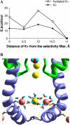
(A) Energy of a K+ ion (♦) and the same ion with the first hydration shell comprising eight water molecules (□) pulled via the Shaker locked-open double mutant Val-474-Cys/Val-476-Cys blocked by two Cd2+ ions. The pulling was accomplished by constraining K+ to the plane, which was translated normally to the pore axis. The abscissa shows the distance (d) between the plane and the K+ ion bound to Thr residues in the selectivity-filter sequence TVGYG. The energy values are partitioned from the structures MC-minimized at each position of the K+-constraining plane. The energies are given relative to the point d = 11 Å. Note that at position d = 10 Å, K+ contributes ∼50% to the energy barrier, and eight water molecules contribute the remaining ∼50%. (B) MC-minimized structure with hydrated K+ constrained at the level of d = 10 Å. In addition to electrostatic repulsion between the closely spaced cations, the system is destabilized by the unfavorable orientation of water molecules, some of which cannot avoid exposure of their hydrogen atoms to Cd2+ ions.

Similar articles

Cited by

References

    1. Doyle, D. A., J. Morais Cabral, R. A. Pfuetzner, A. Kuo, J. M. Gulbis, S. L. Cohen, B. T. Chait, and R. MacKinnon. 1998. The structure of the potassium channel: molecular basis of K+ conduction and selectivity. Science. 280:69–77. - PubMed
    1. Jiang, Y., A. Lee, J. Chen, M. Cadene, B. T. Chait, and R. MacKinnon. 2002. Crystal structure and mechanism of a calcium-gated potassium channel. Nature. 417:515–522. - PubMed
    1. Jiang, Y., A. Lee, J. Chen, V. Ruta, M. Cadene, B. T. Chait, and R. MacKinnon. 2003. X-ray structure of a voltage-dependent K+ channel. Nature. 423:33–41. - PubMed
    1. Jiang, Y., A. Lee, J. Chen, M. Cadene, B. T. Chait, and R. MacKinnon. 2002. The open pore conformation of potassium channels. Nature. 417:523–526. - PubMed
    1. Luzhkov, V. B., J. Nilsson, P. Arhem, and J. Aqvist. 2003. Computational modelling of the open-state Kv 1.5 ion channel block by bupivacaine. Biochim. Biophys. Acta. 1652:35–51. - PubMed

Publication types

LinkOut - more resources