. 2005 May 20;33(9):2887-900.
doi: 10.1093/nar/gki606. Print 2005.

Formation and properties of hairpin and tetraplex structures of guanine-rich regulatory sequences of muscle-specific genes

Affiliations

Formation and properties of hairpin and tetraplex structures of guanine-rich regulatory sequences of muscle-specific genes

Anat Yafe et al. Nucleic Acids Res. .

Abstract

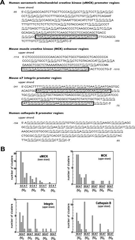
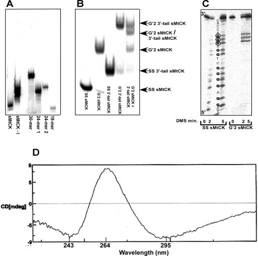
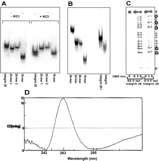
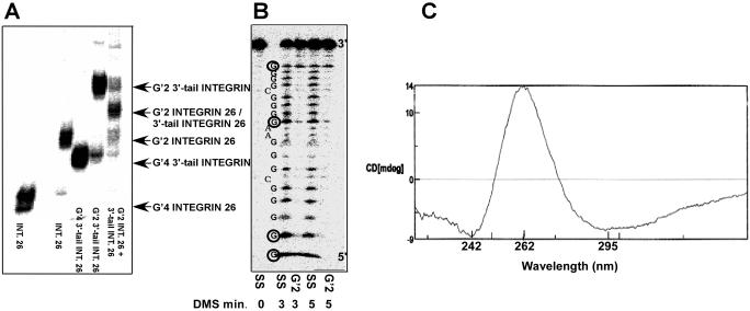
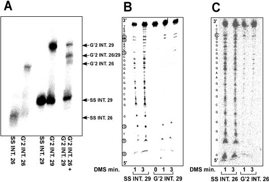
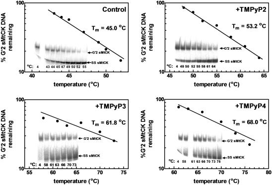
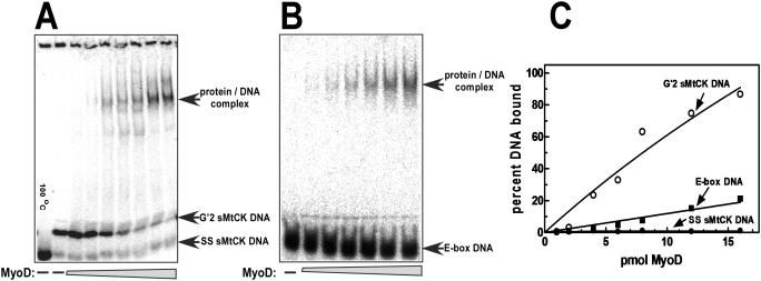
Clustered guanine residues in DNA readily generate hairpin or a variety of tetrahelical structures. The myogenic determination protein MyoD was reported to bind to a tetrahelical structure of guanine-rich enhancer sequence of muscle creatine kinase (MCK) more tightly than to its target E-box motif [K. Walsh and A. Gualberto (1992) J. Biol. Chem., 267, 13714-13718], suggesting that tetraplex structures of regulatory sequences of muscle-specific genes could contribute to transcriptional regulation. In the current study we show that promoter or enhancer sequences of various muscle-specific genes display a disproportionately high incidence of guanine clusters. The sequences derived from the guanine-rich promoter or enhancer regions of three muscle-specific genes, human sarcomeric mitochondrial creatine kinase (sMtCK), mouse MCK and alpha7 integrin formed diverse secondary structures. The sMtCK sequence folded into a hairpin structure; the alpha7 integrin oligonucleotide generated a unimolecular tetraplex; and sequences from all three genes associated to generate bimolecular tetraplexes. Furthermore, two neighboring non-contiguous guanine-rich tracts in the alpha7 integrin promoter region also paired to form a tetraplex structure. We also show that homodimeric MyoD bound bimolecular tetraplex structures of muscle-specific regulatory sequences more efficiently than its target E-box motif. These results are consistent with a role of tetrahelical structures of DNA in the regulation of muscle-specific gene expression.


Figures

Figure 1
Figure 1
High relative incidence of guanine clusters in regulatory regions of four muscle-specific genes. (A) Promoter or enhancer regions of human sMtCK (accession no. M72981), mouse MCK (accession no. M21390), mouse α7 integrin (accession no. U60419) and human cathepsin B (accession no. AF086639) have a high proportion of contiguous guanine residues. Clusters of two or more contiguous guanine residues are marked in boldface and underlined. Sequences of the DNA oligomers used in this work (Table 1) are enclosed in boxes. (B) Occurrence of clusters of two or more contiguous residues of each nucleotide in the muscle-specific regulatory sequences shown in (A). The number of guanine residues is exclusive for each cluster group such that N = 3, for instance, does not include clusters of four or more guanines.
Figure 2
Figure 2
The sMtCK oligonucleotide forms hairpin and G′2 bimolecular tetraplex structures. (A) sMtCK DNA oligomer but not sMtCK-I DNA behaves as a compact structure in the absence of KCl. 5′-32P-labeled sMtCK DNA or sMtCK-I DNA oligomers, each at 0.2 ng/μl in H2O, were boiled for 5 min, rapidly cooled on ice and resolved by non-denaturing electrophoresis through 15% polyacrylamide gel in 0.5× TBE buffer side-by-side with labeled molecular size marker DNA oligomers (see Materials and Methods). (B) sMtCK DNA forms bimolecular complexes. An aliquot of 0.6 μg/μl of 5′-32P-labeled sMtCK or 3′-tail sMtCK DNA oligomer or an equimolar mixture thereof were incubated at 37°C for 16 h in TE buffer, 300 mM KCl and resolved by electrophoresis through a non-denaturing 10% polyacrylamide gel in 0.5× TBE buffer containing 20 mM KCl. Positions of boiled, single-stranded sMtCK or 3′-tail sMtCK DNA oligomers and of their respective slowly migrating G′2 complexes are indicated. Note the presence of a hybrid band of G′2 sMtCK/3′-tail sMtCK DNA in the lane containing an equimolar mixture of the two oligomers. (C) Patterns of methylation protection of single-stranded and G′2 tetraplex sMtCK DNA. 5′-32P-labeled single-stranded sMtCK DNA oligomer or its G′2 bimolecular tetraplex DNA structure were exposed to 1% DMS at 20°C for the specified periods of time. The DMS treated single-stranded and tetraplex DNA structures were hydrolyzed by 2.0 M pyrrolidine as described in Materials and Methods. The position of each nucleotide in the sequence is marked on the phosphor image of the gel and DMS modified guanines in the tetraplex DNA structure are circled. (D) CD spectrum of the sMtCK bimolecular tetraplex DNA.
Figure 3
Figure 3
The integrin 26 DNA oligonucleotide folds into a monomolecular tetraplex structure. (A) Integrin 26 DNA oligomer behaves as a compact structure in the presence of KCl. 5′-32P-labeled integrin 26 DNA oligomer at 2.0 ng/μl in H2O was boiled for 5 min in the absence or presence of 50 mM KCl, rapidly cooled on ice and resolved by a non-denaturing 15% PAGE in 0.5× TBE buffer devoid of or containing 20 mM KCl, as the case be (see Materials and Methods). Shown is a composite of phosphor images of two gels one without and the other containing KCl. (B) Substitution of the guanine residues in integrin 26 DNA by inosines prevents its folding into a compact form. 5′-32P-labeled integrin 26 DNA or integrin 26-I DNA oligomers at 2.0 ng/μl each were boiled in the presence of 50 mM KCl, rapidly cooled and resolved by non-denaturing 15% PAGE in 0.5× TBE buffer, 20 mM KCl, side-by-side with 5′-32P-labeled molecular size marker DNA oligomers. (C) Patterns of methylation protection of single-stranded and monomolecular tetraplex integrin DNA. 5′-32P-labeled single-stranded integrin 26 DNA oligomer or its monomolecular tetraplex form were prepared without or with 50 mM KCl, respectively, as described above. The DNA samples were exposed to 1% DMS and hydrolyzed by 2.0 M pyrrolidine (see Materials and Methods). Shown is a phosphor image of DNA oligomers resolved by electrophoresis in denaturing 12% polyacrylamide gel and 8.0 M urea. The position of each guanine in the sequence is marked in the phosphor image of the gel and DMS modified, pyrrolidine hydrolyzed residues are circled. (D) CD spectrum of integrin 26 monomolecular tetraplex.
Figure 4
Figure 4
The integrin 26 DNA oligonucleotide forms a G′2 bimolecular tetraplex structure. (A) Formation and stoichiometry of bimolecular complexes of integrin 26 DNA. 5′-32P-labeled integrin 26 or 3′-tail integrin 26 DNA oligomers or an equimolar mixture thereof at a final concentration of 0.6 μg/μl were incubated at 37°C for 18 h in TE buffer containing 300 mM KCl and resolved by a non-denaturing 10% PAGE in 0.5× TBE buffer containing 20 mM KCl. Positions of boiled integrin 26 DNA or 3′-tail integrin 26 DNA and their respective slowly migrating G′2 complexes are indicated. Notice the presence of a hybrid band of G′2 integrin 26 DNA/3′-tail integrin 26 DNA in the lane containing an equimolar mixture of the two oligomers. (B) Patterns of methylation protection of single-stranded and G′2 tetraplex integrin 26 DNA. 5′-32P-labeled single-stranded integrin 26 DNA oligomer and its G′2 bimolecular tetraplex form that were prepared as described above were exposed to 1% DMS at 20°C for the specified periods of time, and hydrolyzed by 2.0 M pyrrolidine as described in Materials and Methods and in the legend to Figure 3). DMS modified, pyrrolidine hydrolyzed guanine residues in the tetraplex DNA structure are circled. (C) CD spectrum of G′2 tetraplex integrin 26.
Figure 5
Figure 5
The neighboring integrin 26 and integrin 29 DNA sequences associate to form a G′2 bimolecular tetraplex structure. (A) The integrin 26 and integrin 29 DNA oligomers pair to form bimolecular complexes. 5′-32P-labeled integrin 26 DNA, integrin 29 DNA or their equimolar mixture at a final concentration of 0.2 μg/μl were incubated at 37°C for 18 h in TE buffer that contained 300 mM KCl and resolved by a non-denaturing 10% PAGE in 0.5× TBE buffer containing 20 mM KCl. Positions of boiled single-stranded integrin 26 or integrin 29 DNA and their respective slowly migrating G′2 complexes are indicated. Notice the presence of a hybrid band of G′2 integrin 26 DNA/integrin 29 DNA in the lane containing an equimolar mixture of the two oligomers. (B) Patterns of methylation protection of single-stranded integrin 29 DNA and of the integrin 29 component the G′2 integrin 29 DNA/integrin 26 DNA tetraplex. 5′-32P-labeled single-stranded integrin 29 DNA oligomer and its G′2 bimolecular tetraplex complex with unlabeled integrin 26 DNA were exposed to 0.75% DMS at 20°C for the specified periods of time and hydrolyzed by 2.0 M pyrrolidine as described in Materials and Methods and in the legend to Figure 3. DMS modified and pyrrolidine hydrolyzed guanine residues in the tetraplex DNA structure are circled. (C) Patterns of methylation protection of single-stranded integrin 26 DNA and of the integrin 26 DNA component the G′2 integrin 29 DNA/integrin 26 DNA tetraplex complex. DMS modification, isolation of the treated DNA, pyrrolidine hydrolysis and denaturing gel electrophoresis were as in (B) except that the integrin 26 DNA oligomer was 5′-32P-labeled, whereas the integrin 29 DNA was unlabeled. The circled 3′ guanine residue of integrin 26 DNA in its tetraplex complex with integrin 29 DNA was sensitive to DMS modification.
Figure 6
Figure 6
G′2 sMtCK DNA is stabilized to different extents by three cationic porphyrin isomers. Aliquots of 5′-32P-labeled G′2 tetraplex structure of the sMtCK DNA oligomer were incubated at the specified temperatures for 10 min without or with the indicated cationic porphyrin isomer as detailed in Materials and Methods. Single-stranded and the G′2 tetraplex form of sMtCK DNA were resolved by a non-denaturing 10% PAGE in 0.5× TBE buffer containing 20 mM KCl and their proportions were quantified by phosphor imaging analysis. Shown are semi-logarithmic plots of the fraction of remaining G′2 tetraplex DNA as a function of increasing temperature relative to an initial 100% value determined for G′2 DNA that was maintained at 4°C. Phosphor images of the electrophoretically resolved sMtCK DNA structures are presented in insets within each panel.
Figure 7
Figure 7
MyoD associates preferentially with bimolecular G′2 tetraplex structure of integrin 26 DNA. Recombinant MyoD protein was expressed in E.coli, purified and incubated to form homodimers as described in Materials and Methods. Increasing amounts of the protein were incubated under binding conditions with 0.2 pmol 5′-32P-labeled E-box DNA or G′2 sMtCK DNA and protein–DNA complexes were resolved from free DNA by non-denaturing 4% PAGE in 0.25× TBE buffer containing 10 mM KCl. (A) Binding of MyoD to G′2 sMtCK DNA. Rising amounts of MyoD bound progressively increasing proportions of the G′2 sMtCK DNA whereas the residual hairpin shaped single-stranded DNA did not associate with the protein. (B) Binding of MyoD to E-box DNA. (C) Plots of phosphor imaging quantified results shown in (A and B).

Similar articles

Cited by

References

    1. Simonsson T. G-quadruplex DNA structures—variations on a theme. Biol. Chem. 2001;382:621–628. - PubMed
    1. Shafer R.H., Smirnov I. Biological aspects of DNA/RNA quadruplexes. Biopolymers. 2000;56:209–227. - PubMed
    1. Parkinson G.N., Lee M.P., Neidle S. Crystal structure of parallel quadruplexes from human telomeric DNA. Nature. 2002;417:876–880. - PubMed
    1. Schaffitzel C., Berger I., Postberg J., Hanes J., Lipps H.J., Pluckthun A. In vitro generated antibodies specific for telomeric guanine-quadruplex DNA react with Stylonychia lemnae macronuclei. Proc. Natl Acad. Sci. USA. 2001;98:8572–8577. - PMC - PubMed
    1. Chang C.C., Kuo I.C., Ling I.F., Chen C.T., Chen H.C., Lou P.J., Lin J.J., Chang T.C. Detection of quadruplex DNA structures in human telomeres by a fluorescent carbazole derivative. Anal. Chem. 2004;76:4490–4494. - PubMed

Publication types