. 2005 Jun;138(2):701-14.
doi: 10.1104/pp.105.059774. Epub 2005 May 20.

Loss-of-function mutations of ROOT HAIR DEFECTIVE3 suppress root waving, skewing, and epidermal cell file rotation in Arabidopsis

Collaborators, Affiliations

Loss-of-function mutations of ROOT HAIR DEFECTIVE3 suppress root waving, skewing, and epidermal cell file rotation in Arabidopsis

Christen Y L Yuen et al. Plant Physiol. 2005 Jun.

Abstract

Wild-type Arabidopsis (Arabidopsis thaliana L. Heynh.) roots growing on a tilted surface of impenetrable hard-agar media adopt a wave-like pattern and tend to skew to the right of the gravity vector (when viewed from the back of the plate through the medium). Reversible root-tip rotation often accompanies the clockwise and counterclockwise curves that form each wave. These rotations are manifested by epidermal cell file rotation (CFR) along the root. Loss-of-function alleles of ROOT HAIR DEFECTIVE3 (RHD3), a gene previously implicated in the control of vesicle trafficking between the endoplasmic reticulum and the Golgi compartments, resulted in an almost complete suppression of epidermal CFR, root skewing, and waving on hard-agar surfaces. Several other root hair defective mutants (rhd2-1, rhd4-1, and rhd6-1) did not exhibit dramatic alterations in these root growth behaviors, suggesting that a generalized defect in root hair formation is not responsible for the surface-dependent phenotypes of rhd3. However, similar alterations in root growth behavior were observed in a variety of mutants characterized by defects in cell expansion (cob-1, cob-2, eto1-1, eto2-1, erh2-1, and erh3-1). The erh2-1 and rhd3-1 mutants differed from other anisotropic cell expansion mutants, though, by an inability to respond to low doses of the microtubule-binding drug propyzamide, which normally causes enhanced left-handed CFR and right skewing. We hypothesize that RHD3 may control epidermal CFR, root skewing, and waving on hard-agar surfaces by regulating the traffic of wall- or plasma membrane-associated determinants of anisotropic cell expansion.


Figures
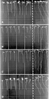
Figure 1.
Figure 1.
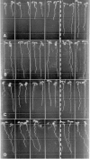
Root-waving phenotypes of wvd6 and rhd3. A, wvd6 (Ws ecotype); B, rhd3-1 (Col ecotype); C, rhd3-2 (Col ecotype); and D, rhd3-3 (No-0 ecotype). Mutant seedlings were plated left of the dotted line, while wild-type seedlings of the same ecotype were plated on the right. Seedlings were grown for 8 to 9 d on 1.5% (w/v) agar-solidified medium, tilted backward 30° from the vertical. Images were taken from the bottom of the plates through the medium.
Figure 2.
Figure 2.
Epidermal CFR phenotypes of wvd6 and rhd3 roots. Seedlings were grown on a tilted agar surface and photographed 9 DAG. Root images were taken either at the tip (A, B, E, and F) or at a position within the mature zone where root hair length was maximal (C and D). A and C, Wild-type Ws; B and D, wvd6; E, wild-type No-0; and F, rhd3-3. Scale bars = 100 μm.
Figure 3.
Figure 3.
wvd6 is caused by a translocation that disrupts RHD3. A, Structure of the wvd6 translocation. The top image represents the arrangement of DNA on the translocated chromosome carrying the chromosome 3 centromere, while the bottom image represents the DNA arrangement on the chromosome carrying the chromosome 1 centromere. The 5′ and 3′ untranslated regions of RHD3 are represented by striped boxes and the coding region of RHD3 by black boxes. Gray boxes are used to represent the predicted first exons of At1g63670 and At1g63680, with arrows indicating the direction of transcription. The gray lines and boxes correspond to DNA originating from chromosome 1, and the black lines and boxes correspond to DNA originating from chromosome 3. Note that the pGKB5 T-DNA inserts are not drawn to scale. The asterisk (*) denotes the truncated T-DNA. The sequences shown represent the various junctions between chromosome 1, chromosome 3, and pGKB5. Sequences in red bold correspond to RHD3 (chromosome 3); sequences in purple italics are from pGKB5; uppercase black sequences are from chromosome 1; and lowercase black sequences are of unknown origin. B, PCR-based confirmation of the wvd6 translocation. Wild-type Ws genomic DNA served as the template for the PCR reactions loaded in lanes 1 and 2, and wvd6 genomic DNA was used for lanes 3 and 4. The primer pair used for lanes 1 and 3 was Chr3F and Chr3R, while Chr1F and Chr3R were used for lanes 2 and 4 (see “Materials and Methods”). Their positions on wild-type chromosomes 1 (top) and 3 (bottom) are depicted by arrows in C, which also shows the translocation breakpoints in the wvd6 mutant. D, From left to right, root growth behavior of 7-d-old wvd6, wvd6 × rhd3-3 (F1), rhd3-3, wvd6 × rhd3-3 (F1), and wild-type No-0 seedlings grown on tilted hard-agar surfaces showing that wvd6 and rhd3-3 do not complement each other.
Figure 4.
Figure 4.
Root-waving phenotypes of root hair defective mutants. A, rhd1-1 (Col ecotype); B, rhd2-1 (Col ecotype); C, rhd4-1 (Col ecotype); and D, rhd6 (Ws ecotype). Mutant seedlings were plated left of the dotted line, while wild-type seedlings of the same ecotype were plated on the right. Seedlings were grown for 8 to 9 d on 1.5% (w/v) agar-solidified medium, tilted backward 30° from the vertical. Images were taken from the bottom of the plates through the medium.
Figure 5.
Figure 5.
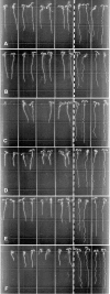
Root-waving phenotypes of root expansion mutants. A, cob-1; B, cob-2; C, eto1-1; D, eto2-1; E, erh2-1; and F, erh3-1. The ecotype background of all of these mutants is Col. Mutant seedlings were plated left of the dotted line, while wild-type Col seedlings were plated on the right. Seedlings were grown for 8 to 9 d on 1.5% (w/v) agar-solidified medium, tilted backward 30° from the vertical. Images were taken from the bottom of the plates through the medium.
Figure 6.
Figure 6.
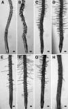
Epidermal CFR phenotypes of root expansion mutants. Seedlings were grown on an agar surface tilted backward by 30° and photographed 9 DAG. All root images were taken at the tip. A, Wild-type Col; B, rhd1-1; C, cob-1; D, cob-2; E, eto1-1; F, eto2-1; G, erh2-1; and H, erh3-1. Scale bars =100 μm.
Figure 7.
Figure 7.
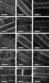
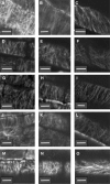
Organization of the cortical microtubule network in wild-type and mutant root tips. Seedlings grown on vertical agar surfaces were subjected to the immunolocalization protocol described in “Materials and Methods” and stained with mouse antitubulin antibodies. Micrographs were taken in the middle of the distal elongation zone (meristem; left column, A, D, G, J, and M), in the middle of the central elongation zone (central column, B, E, H, K, and N), or at the maturing zone where root hairs initiate (right column, C, F, I, L, and O) of wild-type Col (A–C), rhd3-1 (D–F), cob-1 (G–I), erh2-1 (J–L), and eto1-1 (M–O) 5-d-old seedlings. White bars = 10 μm.
Figure 8.
Figure 8.
Organization of the cortical microtubule network in root tips of wild-type and mutant seedlings treated with 3 μm propyzamide. Seedlings grown on vertically oriented plates containing growth media with 3 μm propyzamide were used to analyze organization of root epidermal cell cortical microtubules by whole-mount immunoconfocal microscopy. Micrographs were taken in the middle of the distal elongation zone (left column, A, D, G, J, and M), in the middle of the central elongation zone (central column, B, E, H, K, and N), or at the maturing zone where root hairs initiate (right column, C, F, I, L, and O) of 5-d-old wild-type Col (A–C), rhd3-1 (D–F), cob-1 (G–I), erh2-1 (J–L), and eto1-1 (M–O) seedlings. White bars = 10 μm.

Similar articles

Cited by

References

    1. Andeme-Onzighi C, Sivaguru M, Judy-March J, Baskin T, Driouich A (2002) The reb1-1 mutation of Arabidopsis alters the morphology of trichoblasts, the expression of arabinogalactan-proteins and the organization of cortical microtubules. Planta 215: 949–958 - PubMed
    1. Ausubel F, Brent R, Kingston R, Moore D, Seidman J, Smith J, Struhl K (1994) Current Protcols in Molecular Biology—Updates 1994-2002. John Wiley and Sons, New York
    1. Baskin T, Betzner A, Hoggart R, Cork A, Williamson R (1992) Root morphology mutants in Arabidopsis thaliana. Aust J Plant Physiol 19: 427–437
    1. Baskin T, Wilson J, Cork A, Williamson R (1994) Morphology and microtubule organization in Arabidopsis roots exposed to oryzalin or taxol. Plant Cell Physiol 35: 935–942 - PubMed
    1. Blancaflor E, Masson P (2003) Plant gravitropism. Unraveling the ups and downs of a complex process. Plant Physiol 133: 1677–1690 - PMC - PubMed

Publication types