

doi: 10.1038/nmeth763.
An inducible translocation strategy to rapidly activate and inhibit small GTPase signaling pathways
Affiliations
- PMID: 15908919
- PMCID: PMC3579513
- DOI: 10.1038/nmeth763
Item in Clipboard
An inducible translocation strategy to rapidly activate and inhibit small GTPase signaling pathways
Nat Methods.
2005 Jun.
Abstract
We made substantial advances in the implementation of a rapamycin-triggered heterodimerization strategy. Using molecular engineering of different targeting and enzymatic fusion constructs and a new rapamycin analog, Rho GTPases were directly activated or inactivated on a timescale of seconds, which was followed by pronounced cell morphological changes. As signaling processes often occur within minutes, such rapid perturbations provide a powerful tool to investigate the role, selectivity and timing of Rho GTPase-mediated signaling processes.
Figures
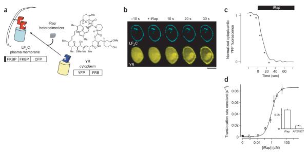
Rapid binding of a cytoplasmic YFP-tagged FRB to a plasma membrane targeted FKBP construct by addition of a rapamycin analog. (a) Schematic representation of the inducible plasma membrane translocation strategy using iRap (solid box; Lyn11). (b) Series of dual confocal images of RBL cells expressing plasma membrane–localized CFP-tagged FKBP (LF2C) and YFP-tagged FRB (YR). iRap triggered a marked translocation of cytosolic YR to the plasma membrane. Live cell dual color measurements were performed on a spinning-disc confocal microscope. For CFP and YFP excitations, we used a helium-cadmium laser and an argon laser, respectively. Fluorescence images were taken every 3 or 5 s during the translocation assay. Scale bar, 10 μm. (c) Normalized fluorescence intensity of YR construct in the cytoplasm before and after addition of iRap. Time points indicated by asterisks correspond to confocal fluorescence images shown in b. (d) Rate constants derived from the membrane translocation timecourse were calculated for measurements taken in the presence of various iRap concentrations. Inset, the rate constants (s−1) of membrane translocation induced by 5 μM iRap and AP21967, respectively. Error bars, s.e.m. (n ≥ 3).
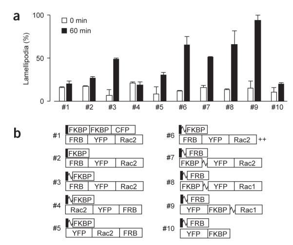
Development of an effective hetero-oligomerization strategy for small GTPase activation. (a) Engineering and testing of FRB, FKBP and Rac constructs to identify suitable heterodimerization pairs for inducing lamellipodia formation. NIH3T3 cells were transfected with each construct pair, stimulated with 5 μM iRap for 60 min, and then fixed and stained with phalloidin. The values shown are numbers of transfected cells with induced lamellipodia. The morphology assay of fixed NIH3T3 cells was performed as described previously14 in the presence of 5 μM iRap at 37 °C. Error bars, s.e.m. (n ≥ 2, 25 ± 2 cells). (b) Schematics of the constructs tested in a (squiggle, flexible linker; ++, polybasic residues GKKKK).
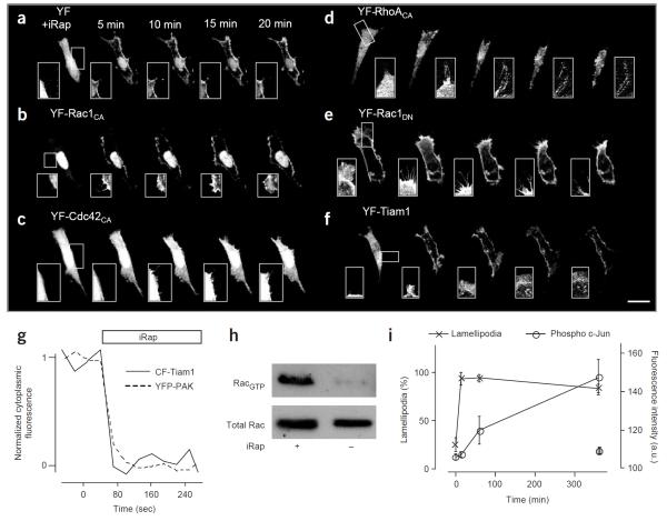
Rapid effects on cell morphology and downstream effectors by heterodimerization. (a–f) Time series of confocal fluorescence images of NIH3T3 cells expressing different pairs of constructs: LDR with YF (a), YF fused to constitutively active Rac1 (b), Cdc42 (c), RhoA (d), Rac1DN (e) or GEF domain of Tiam1 (f). The first image was taken before addition of 5 μM iRap. In the case of Cdc42, neuronal Wiscott-Aldrich syndrome protein (N-WASP) was coexpressed to increase the potential ability of cells to induce filopodia formation. Likewise, cells expressing Rac1DN were pretreated with 10% serum to induce lamellipodia. Inset images highlight a site that shows marked morphological changes (except for the control in a). For the live cell morphology assay, fluorescence images were taken every 30 s with minimal laser power and exposure time to reduce photoinduced damage of the actin cytoskeleton. Scale bar, 20 μm. (g) Kinetic analysis of the translocation of CF-Tiam1 and YFP-PAK to the plasma membrane upon addition of iRap. (h) CF-Tiam1 translocation activates endogenous Rac1. NIH3T3 cells transfected with CF-Tiam1 and LDR were incubated for 30 min in the presence or absence of iRap. Rac1 activity was monitored using pull-down of GTP-bound Rac with a Rac-binding domain coupled to agarose beads. (i) Phosphorylation at Ser63 of c-Jun was detected using nuclear staining with a phosphospecific antibody. The c-Jun phosphorylation level was measured in cells expressing YF-Rac1CA (open circle), and in nontransfected control cells from the same images (gray circle). Lamellipodia formation was evaluated in the same cells and plotted (cross symbol) to compare the timecourse of c-Jun phosphorylation to that of the appearance of morphological changes. Error bars, s.e.m.
References
-
- Takai Y, et al. Physiol. Rev. 2001;81:153–208. - PubMed
-
- Etienne-Manneville S, et al. Nature. 2002;420:629–635. - PubMed
-
- Burridge K, et al. Science. 1999;283:2028–2029. - PubMed
-
- Castellano F, et al. J. Cell Sci. 2000;113:2955–2961. - PubMed
-
- Castellano F, et al. Methods Enzymol. 2000;325:285–295. - PubMed
Publication types
MeSH terms
Substances
Grants and funding
LinkOut - more resources
Full Text Sources
Other Literature Sources
Research Materials
