. 2005 Jun;12(6):539-44.
doi: 10.1038/nsmb938. Epub 2005 May 22.

Internal structure and visualization of transmembrane domains of the RyR1 calcium release channel by cryo-EM

Affiliations

Internal structure and visualization of transmembrane domains of the RyR1 calcium release channel by cryo-EM

Montserrat Samsó et al. Nat Struct Mol Biol. 2005 Jun.

Abstract

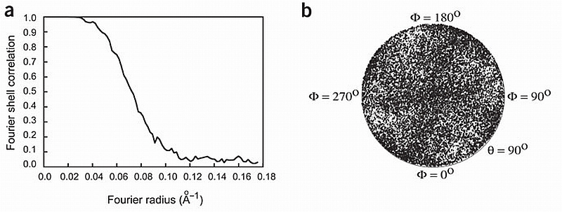
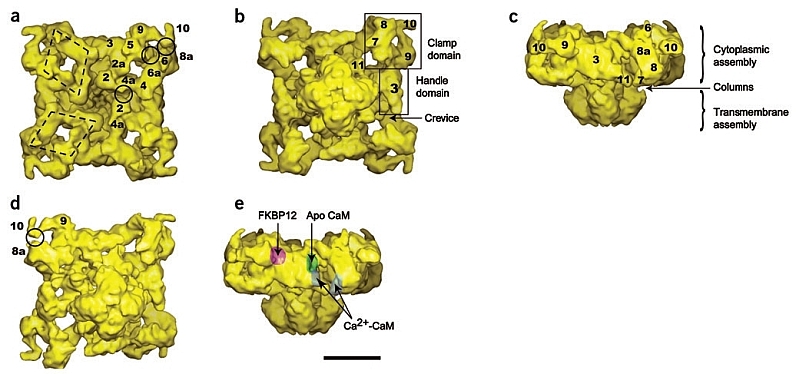
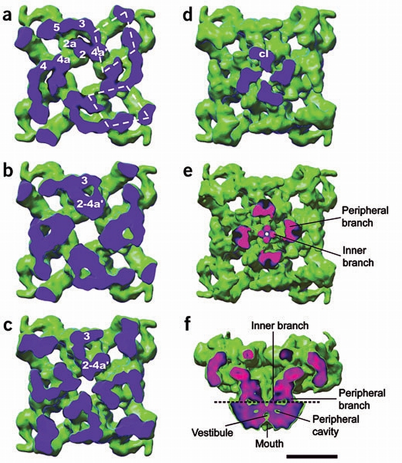
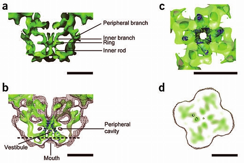
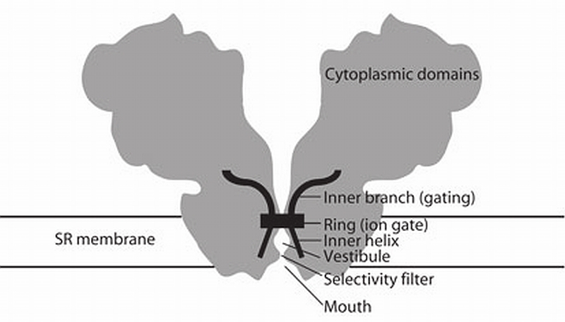
RyR1 is an intracellular calcium channel with a central role in muscle contraction. We obtained a three-dimensional reconstruction of the RyR1 in the closed state at a nominal resolution of approximately 10 A using cryo-EM. The cytoplasmic assembly consists of a series of interconnected tubular structures that merge into four columns that extend into the transmembrane assembly. The transmembrane assembly, which has at least six transmembrane alpha-helices per monomer, has four tilted rods that can be fitted with the inner helices of a closed K(+) channel atomic structure. The rods splay out at the lumenal side and converge into a dense ring at the cytoplasmic side. Another set of four rods emerges from this ring and shapes the inner part of the four columns. The resulting constricted axial structure provides direct continuity between cytoplasmic and transmembrane assemblies, and a possible mechanism for control of channel gating through conformational changes in the cytoplasmic assembly.


Figures

Figure 1
Figure 1
Image processing. (a) Fourier shell correlation curve indicating a resolution of 10.3 Å according to the 0.15 cutoff criterion. (b) Plot of the angular distribution of the particles used in the three‐dimensional reconstruction showing a uniform distribution of orientations of the vitrified RyR.
Figure 2
Figure 2
Isosurface representation of the RyR1 in different views. (a–e) T‐tubule view (a), SR view (b,d), side view (c,e). The SR view in d is slightly tilted with respect to b and contoured at a slightly higher threshold in order to expose the self‐standing structure formed by domains 9‐10 and the separation between domains 10‐8a. In e the approximate positions of the binding sites for FKBP12 (ref. 6), apo CaM and Ca2+‐CaM are indicated with surface shadowing. The clamp, handle, crevice, column (cl) and numeral domain assignations are indicated throughout. The two polygons highlight the rhomboid disposition of the tubular structures defined by domains 2‐4‐5‐6 at their corners. The midpoints of three of the sides of the rhomboids are labeled as domains 2a, 4a and 6a. The full circles indicate areas of close apposition between neighboring domains 2‐4a, 5‐6 and 10‐8a. Scale bar, 10 nm.
Figure 3
Figure 3
Internal structure of the cytoplasmic assembly and the columns. (a–d) Successive sections of the cytoplasmic assembly seen from the SR face show internal mass arrangements and interdomain connections. The sectioning plane is blue. The cytoplasmic domains are interconnected as follows. Below the T‐tubule‐facing surface domains 2 and 4a′ (from adjacent rhomboids) fuse (a). At a lower level, domains 2a and 3 interconnect through a second bridge of density and merge with domains 2‐4a′ (b,c). Therefore each structure formed by the fusion of domains 2‐2a‐3‐4a′ becomes a column of density (d). The four columns of density (cl) in (d) merge into the transmembrane assembly. (e) Section of RyR1 cut along the plane indicated by the dashed line in f seen from the SR lumen, displayed at a slightly higher threshold to show the internal and peripheral regions of the columns. (f) Slice across the side view showing the two branches of each column. The plane of the cut in e,f is color‐coded, with pink corresponding to denser structures and blue to less dense structures. Scale bar, 10 nm.
Figure 4
Figure 4
Substructure of the transmembrane assembly. (a) Central slice of the side view of the transmembrane assembly shown at high threshold to display the rod‐like structures of high density. The two sets of four rods of high density are connected to the ring of high density. (b) Fitting of the inner helices of the K+ channel atomic structure into the central four rods of high density of RyR1. The surface representation is in semitransparent color. A mesh with the surface of RyR1 at a threshold corresponding to RyR1’s molecular mass shows the fitting of the vestibule with the central cavity of the K+ channel. (c) A section of the transmembrane assembly after a 90° rotation shown at the same threshold as in b shows the fitting of the high‐density ring of RyR1 with KcsA’s gate. (d) Slice of the transmembrane assembly of RyR1 perpendicular to the four‐fold symmetry axis with discrete high‐density regions. The position of the slice is indicated by the horizontal dashed line in c. The separation between adjacent high‐density regions is compatible with neighboring α‐helices as shown here with the inner and outer helices of a subunit of the K+ channel in dark blue. Scale bars, 5 nm.
Figure 5
Figure 5
Schematic of the architecture of the RyR. A section across the RyR (gray) is overlaid with the higher‐density, secondary structure—like elements (black). The distinguishable secondary structure elements, and their hypothetical function, are indicated. According to the model proposed here, input from the cytoplasmic assembly would result in a conformational change of the inner branches of the columns. This conformational change would be transmitted directly to the pore inner helices and effectively change the diameter of the gate. An outward pulling of the inner helices in the ring region would open a central path for Ca2+ ions to flow through.

References

    1. Fill M, Copello JA. Ryanodine receptor calcium release channels. Physiol. Rev. 2002;82:893–922. - PubMed
    1. Dulhunty AF, Pouliquin P. What we don’t know about the structure of ryanodine receptor calcium release channels. Clin. Exp. Pharmacol. Physiol. 2003;30:713–723. - PubMed
    1. Wagenknecht T, Samsoó M. Three‐dimensional reconstruction of ryanodine receptors. Front. Biosci. 2002;7:d1464–d1474. - PubMed
    1. Wolf M, Eberhart A, Glossmann H, Striessnig J, Grigorieff N. Visualization of the domain structure of an L‐type Ca2+ channel using electron cryo‐microscopy. J. Mol. Biol. 2003;332:171–182. - PubMed
    1. Paolini C, Protasi F, Franzini‐Armstrong C. The relative position of RyR feet and DHPR tetrads in skeletal muscle. J. Mol. Biol. 2004;342:145–153. - PubMed

Publication types

MeSH terms

Substances