Comparative Study
. 2005 Jul;170(3):1045-62.
doi: 10.1534/genetics.104.040105. Epub 2005 May 23.

In vivo characterization of the nonessential budding yeast anaphase-promoting complex/cyclosome components Swm1p, Mnd2p and Apc9p

Affiliations
Comparative Study

In vivo characterization of the nonessential budding yeast anaphase-promoting complex/cyclosome components Swm1p, Mnd2p and Apc9p

Andrew M Page et al. Genetics. 2005 Jul.

Abstract

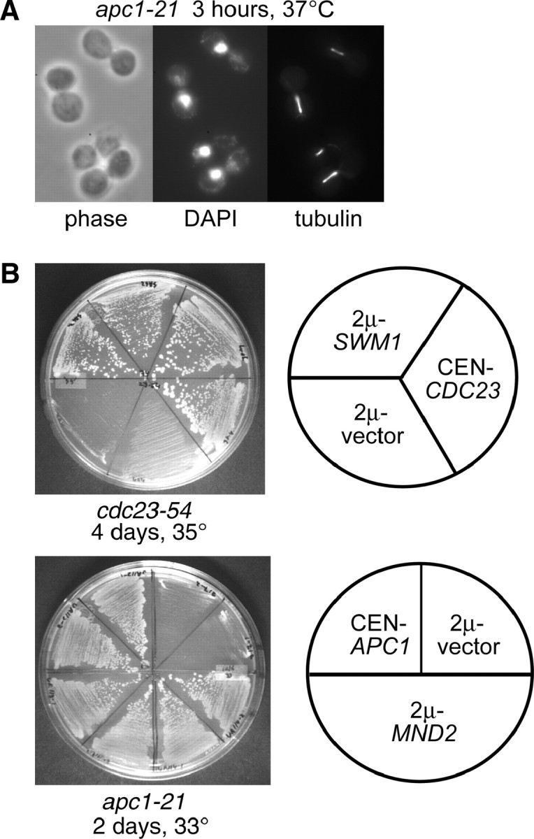
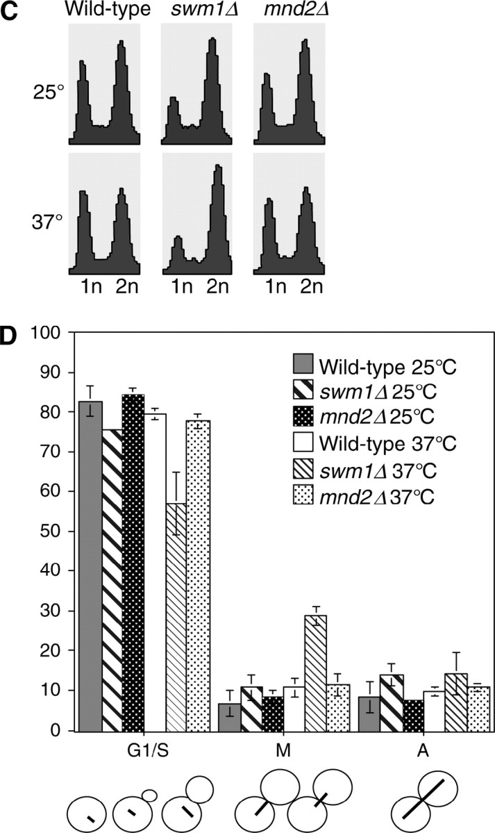
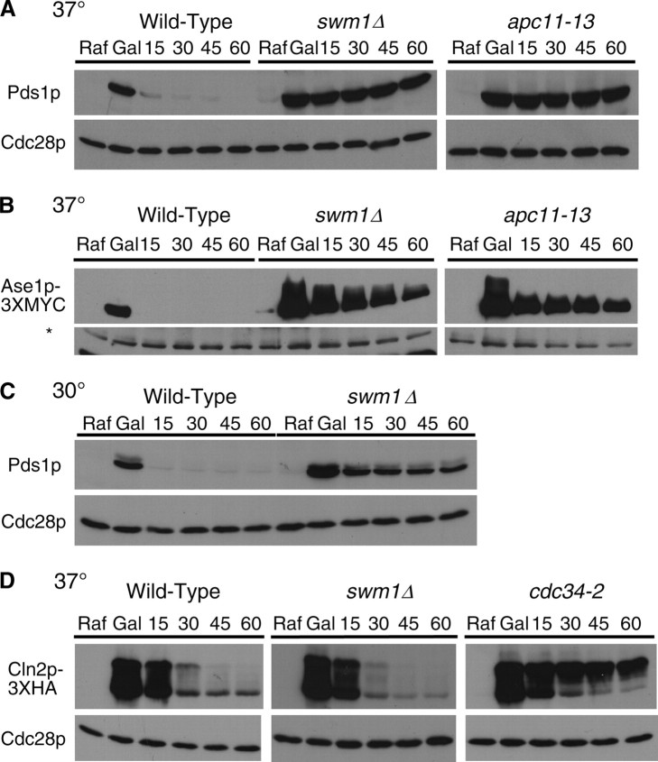
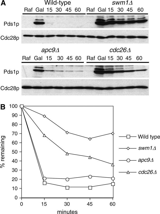
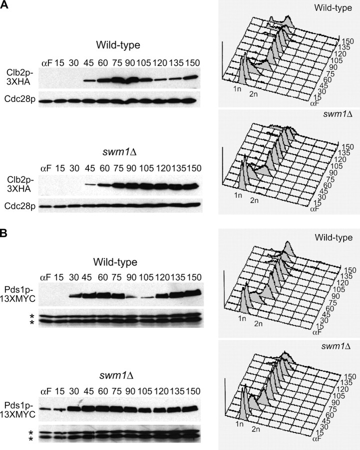
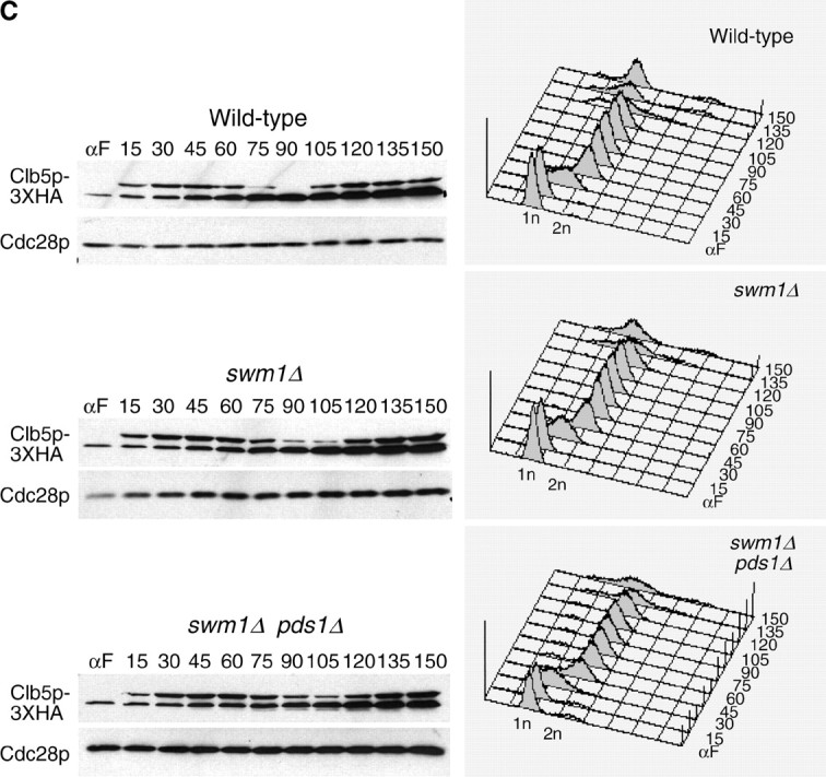
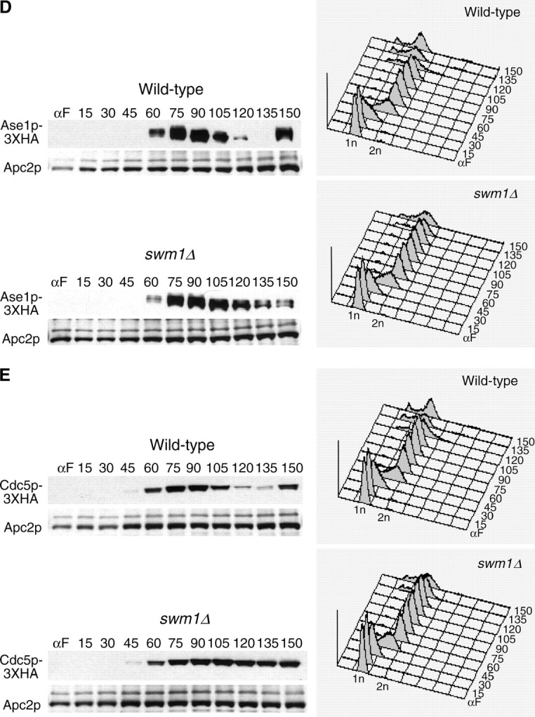
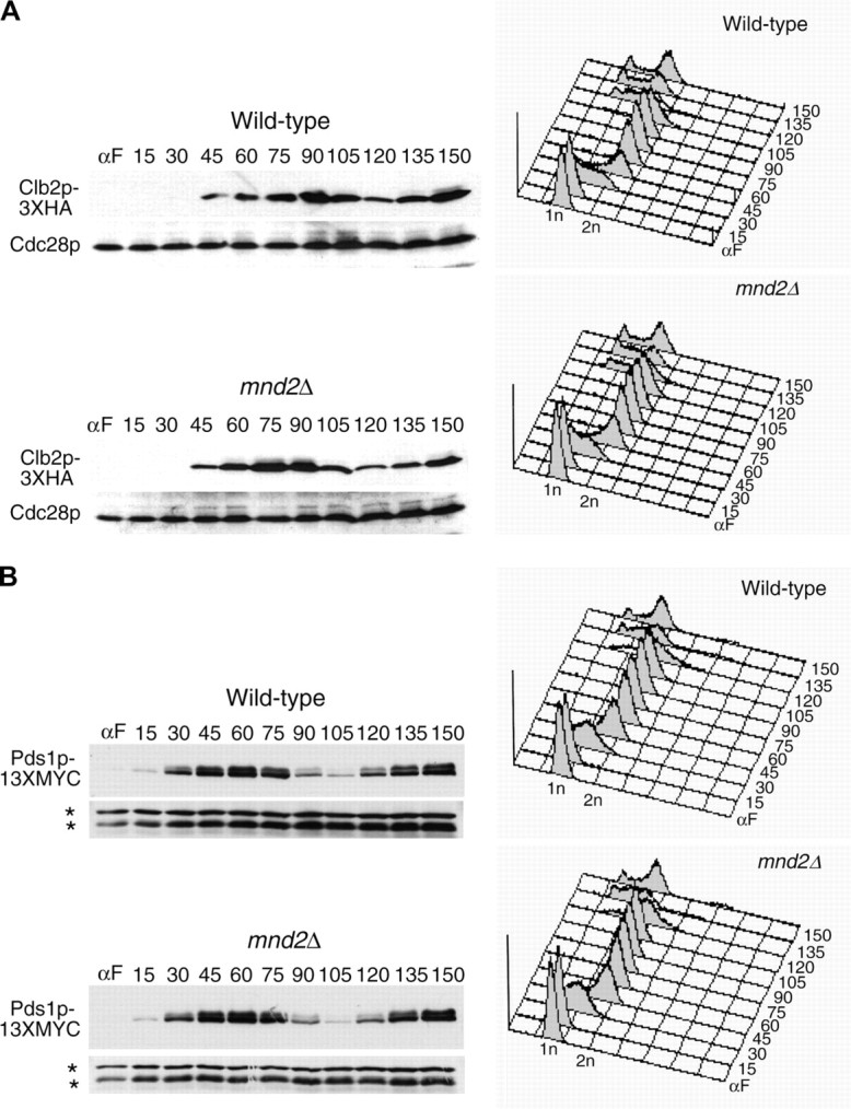
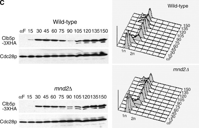
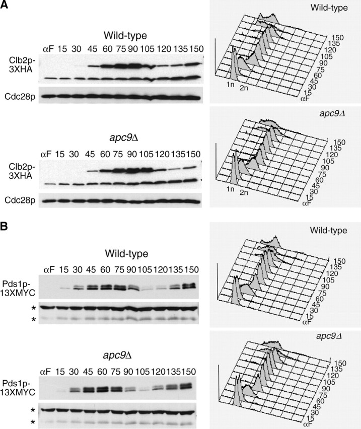
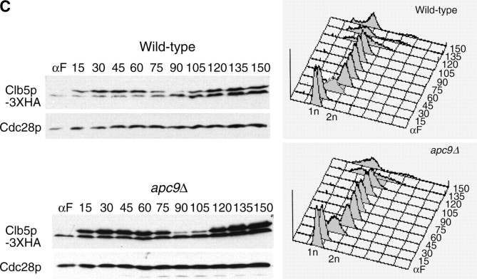
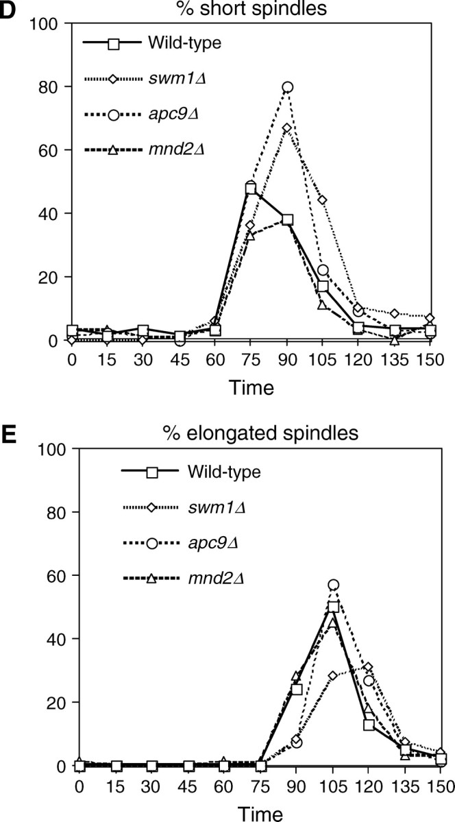
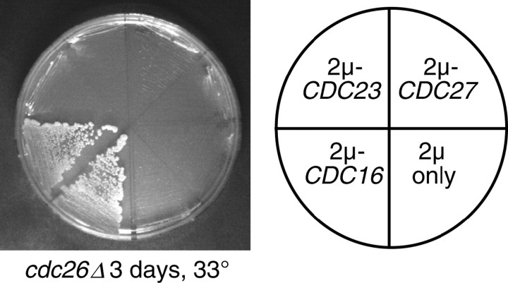
We have examined the in vivo requirement of two recently identified nonessential components of the budding yeast anaphase-promoting complex, Swm1p and Mnd2p, as well as that of the previously identified subunit Apc9p. swm1Delta mutants exhibit synthetic lethality or conditional synthetic lethality with other APC/C subunits and regulators, whereas mnd2Delta mutants are less sensitive to perturbation of the APC/C. swm1Delta mutants, but not mnd2Delta mutants, exhibit defects in APC/C substrate turnover, both during the mitotic cell cycle and in alpha-factor-arrested cells. In contrast, apc9Delta mutants exhibit only minor defects in substrate degradation in alpha-factor-arrested cells. In cycling cells, degradation of Clb2p, but not Pds1p or Clb5p, is delayed in apc9Delta. Our findings suggest that Swm1p is required for full catalytic activity of the APC/C, whereas the requirement of Mnd2p for APC/C function appears to be negligible under standard laboratory conditions. Furthermore, the role of Apc9p in APC/C-dependent ubiquitination may be limited to the proteolysis of a select number of substrates.


Figures

F<sc>igure</sc> 1.—
Figure 1.—
Identification of SWM1 and MND2 as new budding yeast APC/C components. (A) Terminal arrest phenotype of apc1-21. Upon shift to 37° for 3 hr, the majority of apc1-21 cells (YVA1098) arrest with large buds, short mitotic spindles, and DAPI-staining masses at or near the bud neck. (B) SWM1 and MND2 were identified as multicopy suppressors of cdc23-54 and apc1-21, respectively, in independent suppressor screens. A 2μ vector containing the SWM1 open reading frame suppresses the temperature sensitivity of cdc23-54 (YST74) at 35° and a 2μ vector containing MND2 suppresses the temperature sensitivity of apc1-21 (YVA1048) at 33°. (C) Flow cytometry analysis. A wild-type strain (YPH499), swm1Δ (YAP2340), and mnd2Δ (YVA1114) were cultured at 25° and shifted to 37° for 3 hr. Aliquots were harvested and analyzed by flow cytometry or fixed and stained with antitubulin (Yol 1/34) and DAPI. At 25°, swm1Δ has a significant 2N accumulation, whereas the profile of mnd2Δ is similar to that of wild type. Upon shift to 37° for 3 hr, the 2N accumulation of swm1Δ, but not mnd2Δ, is further exacerbated. (D) Upon shift to 37°, the number of large-budded cells with short mitotic spindles at or near the bud neck is approximately threefold higher in swm1Δ compared to wild type.
F<sc>igure</sc> 2.—
Figure 2.—
Swm1p is required for APC/C activity in α-factor-arrested cells. (A) Pds1p wild-type (YAP1146), swm1Δ (YAP1148), or apc11-13 (YAP645) strains bearing pOCF30 (pRS416-GAL1-PDS1) were cultured to midlog phase at 25° in SC-Ura 2% Raf supplemented with additional adenine and tryptophan and arrested for 3.5 hr with 5 μg/ml α-factor at 25°. Cultures were shifted to 37° for 15 min and then induced with 2% galactose for another 30 min at 37°. Two percent glucose and 1 mg/ml cycloheximide was added to repress transcription and translation of the GAL1 promoter, and aliquots were harvested for protein extracts and flow cytometry every 15 min. The “Raf” time point was harvested immediately before shift to 37° and the “Gal” time point was harvested immediately before addition of glucose and cycloheximide. Western blots were probed with anti-Pds1p (C210) and reprobed with anti-Cdc28p as a loading control. (B) Wild-type (YAP1949), swm1Δ (YAP1951), and apc11-13 (YAP1955) cultures all bearing pAP62 (pRS414-GAL1-ASE1-3XMYC) were cultured in SC-Trp 2% Raf supplemented with additional adenine and uracil and arrested as in A. Western blots were probed with anti-MYC (9E10). An anti-Apc2p (BC039) cross-reacting band is used as a loading control. The asterisk represents an unknown anti-Apc2p cross-reacting band used as a loading control. (C) Wild type (YAP1146) and swm1Δ (YAP1148) were cultured to midlog phase in SC-Ura 2% Raf supplemented with additional adenine and tryptophan at 30° and arrested with 5 μg/ml α-factor for 3.5 hr. Cultures were induced with 2% galactose for 30 min and 2% glucose and 1 mg/ml cycloheximide was added to repress GAL1 transcription and translation. Aliquots for cell pellets and flow cytometry were harvested every 15 min and processed as in A. (D) YAP1273 (wild type), YAP1275 (swm1Δ), and YAP1277 (cdc34-2), all bearing pMT634 (pGAL1-CLN2-HA), were grown to midlog phase in SC-Ura 2% Raf supplemented with extra adenine and tryptophan. Cultures were arrested for 3.5 hr in 0.1 m hydroxyurea at 25°, then 2% galactose was added, and cultures were shifted to 37° for 45 min. After galactose induction of Cln2p-3XHA, 2% glucose and 1 mg/ml cycloheximide were added to repress GAL1 transcription and translation and aliquots were harvested for cell pellets and flow cytometry every 15 min. The “Raf” time point was harvested immediately before galactose induction and shift to 37°. Whole-cell lysate was prepared as described in Goh et al. (2000). Western blots were probed with anti-HA (12CA5) and reprobed with anti-Cdc28p as a loading control.
F<sc>igure</sc> 3.—
Figure 3.—
Mnd2p is dispensable for APC/C activity in α-factor-arrested cells. (A) Wild-type (YAP1146), mnd2Δ (YAP2305), and apc1-21 (YAP2841) strains containing pOCF30 were grown to midlog phase at 25° in SC-Ura 2% Raf media supplemented with additional adenine and tryptophan. Cultures were arrested at 25° with 5 μg/ml α-factor for 3.5 hr and then shifted to 37° for 15 min. Two percent galactose was added to induce PDS1 expression for 30 min at 37°. Two percent glucose and 1 mg/ml cycloheximide were then added to repress GAL1 transcription and translation, and aliquots for cell pellets and flow cytometry were harvested every 20 min and processed as in Figure 2A. (B) Wild-type (YAP1949), mnd2Δ (YAP2307), and apc1-21 (YAP2485) strains containing pAP62 were grown to midlog phase at 25° in SC-Trp 2% Raf media supplemented with additional uracil and adenine. Cultures were arrested with 5 μg/ml α-factor at 25° for 3 hr and 45 min and then shifted to 37° for 15 min. Cultures were induced with 2% galactose for 30 min at 37°, and then 2% glucose and 1 mg/ml cycloheximide was added to repress GAL1 transcription and translation. Aliquots for cell pellets and flow cytometry were harvested every 20 min and processed as in Figure 2A. The asterisk represents an unknown anti-Apc2p cross-reacting band used as a loading control.
F<sc>igure</sc> 4.—
Figure 4.—
Deletions of SWM1, APC9, and CDC26 have substantially different APC/C activity in α-factor-arrested cells. (A) Wild-type (YAP1146), swm1Δ (YAP2346), apc9Δ (YAP2771), and cdc26Δ (YAP2775) strains bearing pOCF30 were grown to midlog phase at 25° in SC-Ura 2% Raf supplemented with additional tryptophan and adenine. Cells were arrested with 5 μg/ml α-factor at 25° for 3 hr and 45 min and then shifted to 30° for 15 min. Cultures were induced with 2% galactose for 30 min at 30° and then 2% glucose and 1 mg/ml cycloheximide was added to repress GAL1 transcription and translation. Maintenance of α-factor-arrested cells was verified by flow cytometry (data not shown). Western blots were probed with anti-Pds1p (C210, D. Koshland) and reprobed with anti-Cdc28p as a loading control. (B) Pds1p levels were quantified on a Bio-Rad (Richmond, CA) Fluor-S multi-imager.
F<sc>igure</sc> 5.—
Figure 5.—
Deletion of SWM1 affects APC/C substrate turnover during the mitotic cell cycle. (A) swm1Δ is defective in Clb2p-3XHA degradation at the permissive temperature. Wild-type (YAP1543) and swm1Δ (YAP2410) strains containing CLB2-3XHA were grown to midlog phase in 50 ml YEPD, washed twice with ddH2O, and resuspended in 50 ml YEPD with 5 μg/ml α-factor. Cultures were arrested for 135–150 min at 30°, with a booster dose of 3.75 μg/mLl α-factor added after 90 min. Cells were then harvested and washed twice with ddH2O and resuspended in 30 ml YEPD lacking α-factor. Cultures were standardized to similar density and released into the cell cycle at 25°. Every 15 min, 1 ml of each culture was harvested for cell pellets and 1 ml was harvested for flow cytometry. Cell pellet aliquots were washed with 1 ml ice-cold ddH2O and immediately frozen at −80° until extracts were prepared. Whole-cell lysate was prepared as described in Goh et al. (2000). Western blots were probed with anti-HA (12CA5) and reprobed with anti-Cdc28p as a loading control. (B) Pds1p-13XMYC is partially stabilized in swm1Δ. Wild-type (YAP1232) and swm1Δ (YAP2821) strains containing PDS1-13XMYC were arrested and released into the cell cycle as in A. Western blots were probed with anti-MYC (9E10) and reprobed with anti-Cdc28p. The asterisks represent anti-Cdc28p cross-reacting bands used as a loading control. (C) Delay of Clb5p-3XHA degradation in swm1Δ does not depend on Pds1p. Wild-type (YAP1294), swm1Δ (YAP2827), and swm1Δ pds1Δ (YAP2873) strains containing CLB5-3XHA were arrested and released into the cell cycle as described in A. (D) Ase1p-3XHA is partially stabilized in swm1Δ. Wild-type (YAP1646) and swm1Δ (YAP2861) strains containing ASE1-3XHA were arrested and released into the cell cycle as described in A. (E) Cdc5p-3XHA degradation is defective in swm1Δ. Wild type (YAP2004) and swm1Δ (YAP2864), both containing CDC5-3XHA, were arrested and released into the cell cycle as described in A.
F<sc>igure</sc> 6.—
Figure 6.—
Deletion of MND2 does not affect degradation of APC/C substrates during the mitotic cell cycle. CLB2-3XHA (YAP1543) and CLB2-3XHA mnd2Δ (YAP2419) (A), PDS1-13XMYC (YAP1232) and PDS1-13XMYC mnd2Δ (YAP2824) (B), and CLB5-3XHA (YAP1294) and CLB5-3XHA mnd2Δ (YAP2830) (C) were arrested with α-factor and released into the cell cycle as described in Figure 5A. Deletion of MND2 did not substantially affect the timing of Clb2p-3XHA, Pds1p-13XMYC, or Clb5p-3XHA degradation. Asterisks in B represent anti-Cdc28p cross-reacting bands used as a loading control.
F<sc>igure</sc> 7.—
Figure 7.—
APC/C substrate degradation in apc9Δ. CLB2-3XHA (YAP1543) and CLB2-3XHA apc9Δ (YAP2934) (A), PDS1-13XMYC (YAP1232) and PDS1-13XMYC apc9Δ (YAP2937) (B), and CLB5-3XHA and CLB5-3XHA apc9Δ (YAP2941) (C) were arrested with α-factor and released into the cell cycle as described in Figure 5A. apc9Δ delayed the onset of Clb2p-3XHA proteolysis but did not significantly affect the timing of PDS1-13XMYC and Clb5p-3XHA degradation. Asterisks in B represent anti-Cdc28p cross-reacting bands. (D and E) Wild-type (YPH499), swm1Δ (YAP2340), apc9Δ (YAP2930), and mnd2Δ (YVA1114) were grown to midlog phase in YEPD, arrested with α-factor, and released into the cell cycle as described in Figure 5A. Samples were harvested and fixed for immunofluorescence and stained with antitubulin (Yol 1/34) and DAPI. Short and long mitotic spindles were scored for each time point (n = 100).
F<sc>igure</sc> 8.—
Figure 8.—
Suppression of APC/C double mutants by overexpression of specific TPR proteins. Overexpression of CDC16 suppresses the temperature sensitivity of cdc26Δ. cdc26Δ::KanMX (YVA435) was transformed with 2μ plasmids expressing a single APC/C component or regulator or with empty vector only. Individual transformants were outgrown on selective media at 25° and then applied to selective media at the nonpermissive temperature.

Similar articles

Cited by

References

    1. Adames, N. R., J. R. Oberle and J. A. Cooper, 2001. The surveillance mechanism of the spindle position checkpoint in yeast. J. Cell Biol. 153 159–168. - PMC - PubMed
    1. Amon, A., 2001. Together until separin do us part. Nat. Cell Biol. 3 E12–E14. - PubMed
    1. Amon, A., S. Irniger and K. Nasmyth, 1994. Closing the cell cycle circle in yeast: G2 cyclin proteolysis initiated at mitosis persists until the activation of G1 cyclins in the next cycle. Cell 77 1037–1050. - PubMed
    1. Bachmair, A., D. Finley and A. Varshavsky, 1986. In vivo half-life of a protein is a function of its amino-terminal residue. Science 234 179–186. - PubMed
    1. Bloecher, A., G. M. Venturi and K. Tatchell, 2000. Anaphase spindle position is monitored by the BUB2 checkpoint. Nat. Cell Biol. 2 556–558. - PubMed

Publication types

MeSH terms