Comparative Study
. 2005 May 31;102(22):8006-11.
doi: 10.1073/pnas.0503005102. Epub 2005 May 23.

ExsE, a secreted regulator of type III secretion genes in Pseudomonas aeruginosa

Affiliations
Comparative Study

ExsE, a secreted regulator of type III secretion genes in Pseudomonas aeruginosa

Arne Rietsch et al. Proc Natl Acad Sci U S A. .

Abstract

Type III secretion systems are toxin delivery systems that are present in a large number of pathogens. A hallmark of all type III secretion systems studied to date is that expression of one or more of their components is induced upon cell contact. It has been proposed that this induction is controlled by a negative regulator that is itself secreted by means of the type III secretion machinery. Although candidate proteins for this negative regulator have been proposed in a number of systems, for the most part, a direct demonstration of their role in regulation is lacking. Here, we report the discovery of ExsE, a negative regulator of type III secretion gene expression in Pseudomonas aeruginosa. Deletion of exsE deregulates expression of the type III secretion genes. We provide evidence that ExsE is itself secreted by means of the type III secretion machinery and physically interacts with ExsC, a positive regulator of the type III secretion regulon. Taken together, these data demonstrate that ExsE is the secreted negative regulator that couples triggering of the type III secretion machinery to induction of the type III secretion genes.


Figures

Fig. 1.
Fig. 1.
Genetic organization of the type III secretion regulatory locus. Promoters are indicated by arrows. All genes in this chromosomal fragment are depicted as solid black arrows, except exsE, which is striped. Numbers below the fragment indicate size in base pairs.
Fig. 2.
Fig. 2.
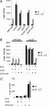
ExsE regulates exoS expression. Expression of a lacZ reporter gene inserted at the exoS locus was monitored by β-galactosidase assay. Cells were grown in the presence (black bars) or absence (striped bars) of calcium. (A) Effect of deletion mutations in the regulatory locus and structural components of the secretion machinery on exoS expression; the genes that were deleted are indicated underneath the respective bars. exsE-myc5 indicates a strain in which exsE has been fused to five copies of the Myc epitope tag. (B) Complementation analysis. Relevant strain genotypes are indicated above the graph. Plasmid information and whether cultures were induced with isopropyl β-d-thiogalactoside (IPTG) (100 μM final concentration) are indicated below the graph. (C) Complementation of an ΔexsC ΔexsE double null mutant strain. Plasmid information and whether cultures were induced with IPTG (250 μM final concentration) are indicated below the graph. β-Galactosidase activity is given in Miller units (35).
Fig. 3.
Fig. 3.
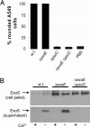
An exsE mutant is secretion-competent. (A) Cytotoxicity was assayed by determining the percentage of rounded A549 cells after 4 h of coincubation with WT PAK or the indicated deletion mutants. (B) ExoS in cell pellets or supernatant fractions was identified by Western blot. The location of the band corresponding to ExoS is indicated by an arrow. Genotypes of the strains assayed are indicated above the gel. Presence or absence of calcium in the medium is indicated below the gel.
Fig. 4.
Fig. 4.
ExsE is secreted in a type III-dependent manner. A Myc5-tagged version of ExsE was detected in supernatant or cell pellet fractions of induced or uninduced bacteria. The relevant genotype is indicated above the gel. The presence or absence of calcium in the culture media is noted below the gel. A WT strain without the Myc5-tagged ExsE was run as a specificity control for the tagged protein.
Fig. 5.
Fig. 5.
ExsE and ExsC interact. (A) The interaction between ExsE and ExsC was monitored by two-hybrid analysis. Increasing concentrations of isopropyl β-d-thiogalactoside (IPTG) with which expression of the fusion partners was induced are indicated on the x axis. β-Galactosidase activity is noted on the y axis. The fusion proteins being assayed are indicated to the right. (B) Copurification of ExsE-Myc5 with either TAP-tagged NusA or TAP-tagged ExsC. The presence of ExsE was monitored by Western blot. The fraction assayed is indicated above the gel. The ExsE-Myc5 band is indicated by an arrow. The TAP-tagged fusion protein present in each lane is indicated below the gel.
Fig. 6.
Fig. 6.
A PcrV mutant secretes ExoS constitutively. The presence of ExoS in TCA-precipitated culture supernatants was monitored by Western blot. The presence or absence of calcium in the medium is noted below the gel. The growth phase of the bacteria is noted above the gel, as is the relevant genotype of each strain assayed.
Fig. 7.
Fig. 7.
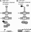
A model for induction of the type III secretion regulon. (A and B) All proteins except ExsE (E) and PcrV (V) are indicated by name. The type III secretion machinery is depicted spanning the inner membrane (IM) and outer membrane (OM). Expression of exoS under inducing conditions is indicated by a wavy arrow.

References

    1. Nemunaitis, J., Buckner, C. D., Dorsey, K. S., Willis, D., Meyer, W. & Appelbaum, F. (1998) Am. J. Clin. Oncol. 21, 341–346. - PubMed
    1. Velasco, E., Thuler, L. C., Martins, C. A., Dias, L. M. & Goncalves, V. M. (1997) Am. J. Infect. Control 25, 458–462. - PubMed
    1. Carratala, J., Roson, B., Fernandez-Sevilla, A., Alcaide, F. & Gudiol, F. (1998) Arch. Intern. Med. 158, 868–872. - PubMed
    1. Cheng, K. H., Leung, S. L., Hoekman, H. W., Beekhuis, W. H., Mulder, P. G., Geerards, A. J. & Kijlstra, A. (1999) Lancet 354, 181–185. - PubMed
    1. Beers, S. L. & Abramo, T. J. (2004) Pediatr. Emerg. Care 20, 250–256. - PubMed

Publication types

MeSH terms