. 1979 Oct;18(2):297-307.
doi: 10.1016/0092-8674(79)90049-7.

Interaction of int protein with specific sites on lambda att DNA

Interaction of int protein with specific sites on lambda att DNA

W Ross et al. Cell. 1979 Oct.

Abstract

We have studied the interaction of highly purified Int protein with DNA restriction fragments from the lambda phage attachment site (attP) region. Two different DNA sequences are protected by bound Int protein against partial digestion by either pancreatic DNAase or neocarzinostatin. One Int binding site includes the 15 bp common core sequence (the crossover region for site-specific recombination) plus several bases of sequence adjoining the core in both the P and P' arms. The second Int-protected site occurs 70 bp to the right of the common core in the P' arm, just at the distal end of the sequence encoding Int protein. The two Int binding sites are of comparable size, 30-35 bp, but do not share any extensive sequence homology. The interaction of Int with the two sites is distinctly different, as defined by the observation that only the site in the P' arm and not the site at the common core region is protected by Int in the face of challenge by the polyanion heparin. Restriction fragments containing DNA from the bacterial attachment site (attB) region exhibit a different pattern of interaction with Int. In the absence of heparin, a smaller (15 bp) sequence, which includes the left half of the common core region and the common core-B arm juncture, is protected against nuclease digestion by Int protein. No sequences from this region are protected by Int in the presence of heparin.


Figures

Figure 1
Figure 1. DNA Restriction Fragments Used in the Footprinting Experiments
The locations of known restriction enzyme recognition sites are indicated for the POP′ (attP) and BOB′ (attB) regions studied in this work. The attachment site sequences are numbered with 0 as the central base of the 15 bp common core sequence (■); negative numbers proceed leftward from 0 into P or B arm sequences and positive numbers extend rightward from 0 into P′ or B′ arm sequences (Landy and Ross, 1977). The position of sequence encoding the carboxy terminal end of the Int protein is indicated ( formula image
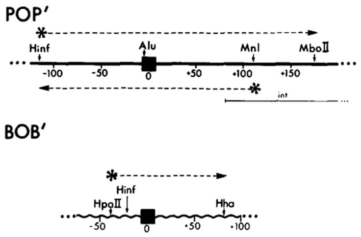
) (see Figure 5 and Discussion). The bottom strand corresponds to the Int message. Two restriction fragments from the attP region were used: one, Hinf I (−114)-Mbo II (+173), was labeled with 32P (*) at the Hinf I site at the 5′ end of the top strand (* ‒ ‒ →); the second, Hinf I (−114)-Mnl I (+115) (← ‒ ‒ *), was labeled at the Mnl I site at the 5′ end of the bottom strand. The attB restriction fragment Hpa II (−43)-Hha I (+80) was labeled at the Hpa II site at the 5′ end of the top strand (see Experimental Procedures).
Figure 2
Figure 2. Pancreatic DNAase Footprints of Int on the Bottom Strand of a Restriction Fragment from the attP Region
The fragment Hinf I (−114)-Mnl I ( + 115) is labeled with 32P at the Mnl I end (see Figure 1). Lanes 1 and 2 in each panel contain DNA sequence markers. A + G and C + T, prepared by the method of Maxam and Gilbert(see Experimental Procedures). See Figure 1 legend for numbering of sequences. The sequence is presented in Figure 5 and in Landy and Ross (1977). In (A), the DNA in samples 3 and 4 was digested with DNAase in the absence of Int, either with (lane 4) or without (lane 3) the prior addition of 5 μg/ml heparin. DNA samples in lanes 5–8 were incubated with either a low concentration of Int (1.25 μg/ml, lanes 5 and 6) or a high concentration of Int (10 μg/ml, lanes 7 and 8). Int-DNA complexes in samples 6 and 8 were also challenged with 5 μg/ml heparin prior to DNAase digestion (see Experimental Procedures). In (B), where the samples were electrophoresed for longer times to resolve the common core region better, lane 3 contains a control sample(DNAase digested in the absence of Int) while samples 4 and 5 were incubated with 1.25 (4) or 2.5 μg/ml (5) of Int (low Int) prior to DNAase digestion. No heparin was used in any of the samples in (B). DNA sequences protected from DNAase digestion are bracketed and the sequence coordinates of the boundaries of the protection are indicated beside the brackets. Two coordinates for a protection boundary reflect uncertainty as to the precise position of the boundary (see Results and Figure 5). Pancreatic DNAase digestion products have slightly different electrophoretic mobility than sequence markers (see Results).
Figure 3
Figure 3. Neocarzinostatin Footprints of Int on the Bottom Strand of a Restriction Fragment from the attP Region
The restriction fragment Hinf I-Mnl I is the same as that used in Figure 2 (see Figure 1 and Experimental Procedures). Chemical sequence markers, prepared by the method of Maxam and Gilbert, are found in lanes 1 and 2 in each panel. Numbering of the sequence is as described in the legend to Figure 1. Samples in (A) and (C) are identical; the gel in (C) was electrophoresed for a longer time to resolve the common core region better. Samples in lanes 3 and 4 of (A) and (C) were digested with NCS in the absence of Int, and either with (lanes 4) or without (lanes 3) the prior addition of 5 μg/ml heparin. Samples in lanes 5 and 6 were incubated with 1.25 μg/ml Int (low Int), then either challenged with heparin (lanes 6) or not challenged (lanes 5) prior to NCS digestion. (B) illustrates NCS digestion profiles of samples incubated with a high concentration of Int (10 μg/ml), with (lane 5) or without (lane 4) subsequent challenge with heparin. Brackets identify the regions of sequence which are protected by Int from NCS digestion, and the sequence coordinates indicate the boundaries of protection. See Figure 5 for sequence detail. Arrows (←) within brackets indicate less well protected bases in the common core sequence (see Results).
Figure 4
Figure 4. Neocarzinostatin Footprints of Int on the Top Strand of a Restriction Fragment from the attP Region
The fragment used was Hinf I (−114)-Mbo II (+173), labeled at the Hinf I end (see Figure 1). C + T and A + G chemical sequence markers (see legend to Figure 2) are in lanes 1 and 2. A control sample, digested with NCS in the absence of Int and heparin, is in lane 4. (Heparin does not influence the extent or specificity of NCS cleavage; see Figure 3A, lanes 3 and 4.) In lane 3, the DNA sample was incubated with 1.25 μg/ml Int (low Int) and not challenged with heparin prior to NCS digestion. DNA in samples 5 and 6 was incubated with a high concentration of Int (10 μg/ml), with (lane 6) or without (lane 5) challenge by heparin prior to NCS digestion. Following the digestion, samples 5 and 6 were passed through nitrocellulose filters, and the DNA retained on the filters was eluted off and applied to the gel (see Experimental Procedures). Brackets designate sequence protected from NCS cleavage by Int (see Figure 5). The boundaries of protection were determined from a gel electrophoresed for a longer time (not shown). The arrow (←) in the lower bracket indicates less well protected bases in the common core protected region.
Figure 5
Figure 5. Two Sequences from the attP Region Protected by Int from Digestion with Either Neocarzinostatin or Pancreatic DNAase
The top sequence includes the common core protected region; letters in bold type are the 15 bases of the common core sequence. The bottom sequence includes the protected region in the P′ arm. See Figure 1 legend for sequence numbering. Solid bars (■) indicate sequences protected from NCS digestion; open bars (□) indicate sequences protected from pancreatic DNAase digestion. Bars are drawn above the sequence when the data were obtained from a fragment labeled in the top strand, and below the sequence when the fragment used was labeled in the bottom strand. A base is designated as being protected from pancreatic DNAase if its 5′ phosphodiester linkage is protected. (●) or (○) indicate bases whose extent of protection from NCS or DNAase digestion is not known, due to failure of the nuclease to cut the base in an unprotected control sample (see Results). Two tandem stop codons (5′...UAAUGA...) which define the carboxy terminal end of the Int gene are indicated on the bottom strand (Int message strand; see Discussion). A 10 base sequence which is also found as part of an inverted repeat structure at the ends of Tn10 is indicated in the P′ arm protected region (Kleckner, 1979). A sequence homology between the common core and P′ arm protected regions is indicated with a line (—) between the two strands of the sequences. ( formula image
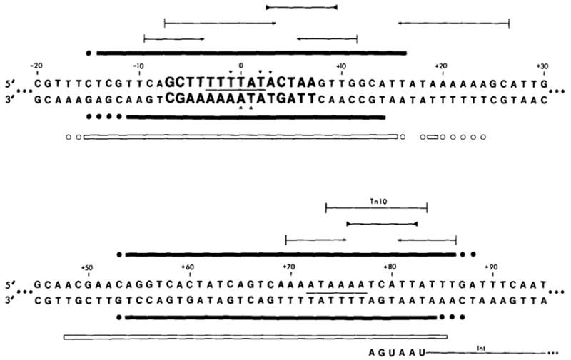
) Inverted repeat; ( formula image) palindromic sequences. Less well protected bases in NCS experiments with the top strand (▼); or the bottom strand (▲) (see Results).
Figure 6
Figure 6. Pancreatic DNAase (A), Neocarzinostatin (B) and Micrococcal Nuclease (C) Footprints of Int on the Top Strand of a Restriction Fragment from the attB Region
The restriction fragment Hpa II (−43)-Hha I (+80) is labeled at the Hpa II end (see Figure 1). Chemical sequence markers, as described in the legend to Figure 2 and Experimental Procedures, are in lanes 1 and 2. In (A) and (C), DNA samples were incubated without Int (lane 3) or with varying concentrations of Int [1.25 μg/ml (1X) (lane 4), 2.5 μg/ml (2X) (lane 5) or 5 μg/ml (4X) (lane 6, panel A)] prior to digestion with the specified nuclease. No heparin was used in these experiments. In (B), DNA samples were incubated in the absence of Int (lanes 3 and 4) or in the presence of 1.25 μg/ml (low) Int (lanes 5 and 6). Incubated samples in lanes 4 and 6 were challenged with heparin prior to NCS digestion. Brackets identify regions protected by Int from digestion. Protected sequences are illustrated in Figure 7. The boundaries of the region at +38 to + 50–52 were determined from a second gel (not shown).
Figure 7
Figure 7. Two Sequences from the attB Region Protected by Int from Nuclease Digestion
The top sequence includes the 15 base common core sequence (bold type). The bottom sequence is from the B′ arm. See the legend to Figure 1 for sequence numbering. (■) Sequences protected from NCS; (□) sequences protected from pancreatic DNAase; ( formula image
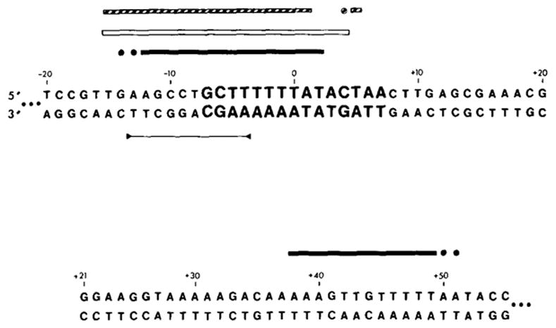
) sequences protected from micrococcal nuclease. All experiments were carried out with a fragment labeled in the top strand (see Figures 1 and 6). (●), (○) or ( formula image) Bases whose extent of protection is not known; see legend to Figure 5 and Results. ( formula image) A palindromic sequence.

References

    1. Bernardi A, Gaillard C, Bernardi G. The specificity of five DNAases as studied by the analysis of 5′-terminal doublets. Eur J Biochem. 1975;52:451–457. - PubMed
    1. Bidwell K, Landy A. Structural features of λ site-specific recombination at a secondary att site in ga/T. Cell. 1979;16:397–406. - PubMed
    1. Campbell A, Heffernan L, Hu S-L, Szybalski W. The integrase promoter of bacteriophage lambda. In: Bukhari AI, Shapiro JA, Adhya SL, editors. DNA Insertion Elements, Plasmids, and Episomes. New York: Cold Spring Harbor Laboratory; 1977. pp. 375–379.
    1. Chamberlin MJ. RNA polymerase—an overview. In: Losick R, Chamberlin M, editors. RNA Polymerase. New York: Cold Spring Harbor Laboratory; 1976. pp. 35–37.
    1. Christie GE, Platt T. A secondary attachment site for bacteriophage λ in trpC of E. coli. Cell. 1979;16:407–413. - PubMed

Publication types

Associated data

LinkOut - more resources