. 2005 May 24:5:39.
doi: 10.1186/1471-2334-5-39.

A sensitive flow cytometric methodology for studying the binding of L. chagasi to canine peritoneal macrophages

Affiliations

A sensitive flow cytometric methodology for studying the binding of L. chagasi to canine peritoneal macrophages

Ricardo Gonçalves et al. BMC Infect Dis. .

Abstract

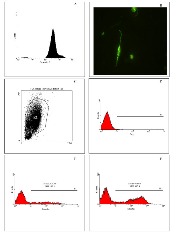
Background: The Leishmania promastigote-macrophage interaction occurs through the association of multiple receptors on the biological membrane surfaces. The success of the parasite infection is dramatically dependent on this early interaction in the vertebrate host, which permits or not the development of the disease. In this study we propose a novel methodology using flow cytometry to study this interaction, and compare it with a previously described "in vitro" binding assay.

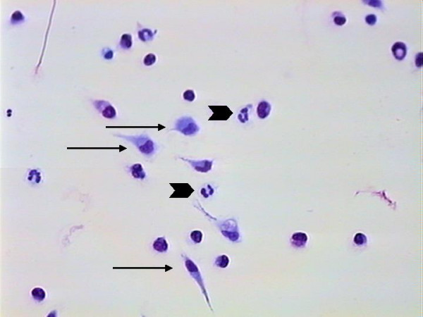
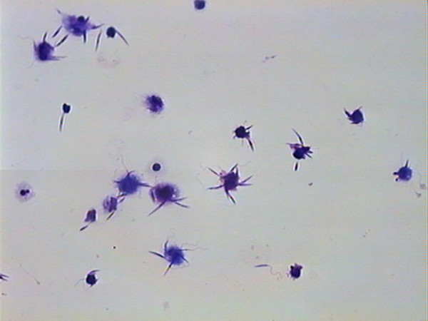
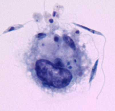
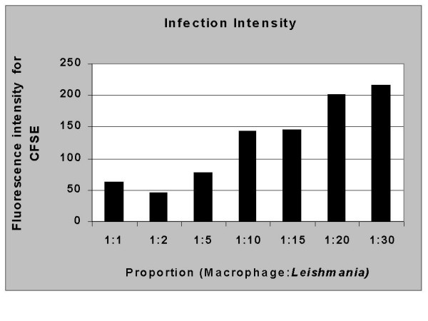
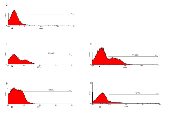
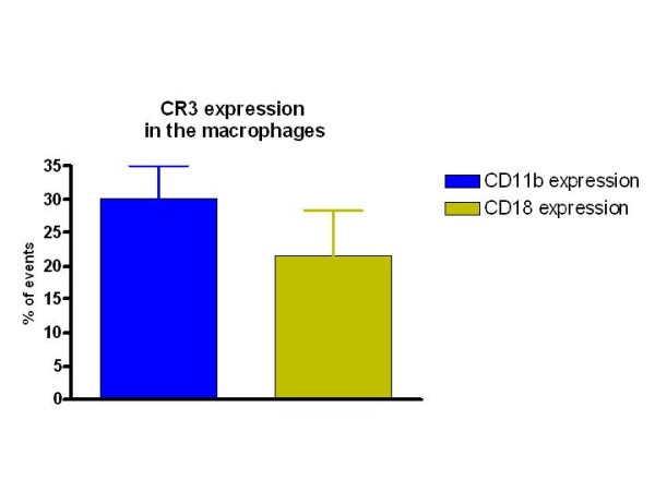
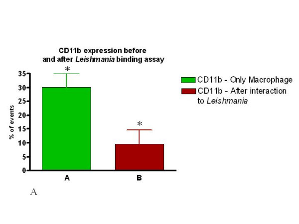
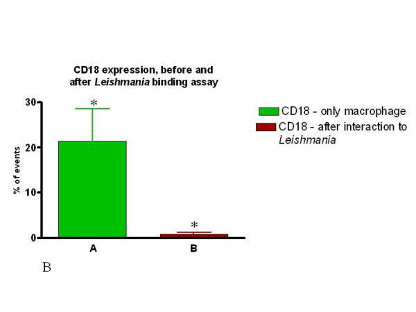
Methods: To study parasite-macrophage interaction, peritoneal macrophages were obtained from 4 dogs and adjusted to 3 x 10(6) cells/mL. Leishmania (Leishmania) chagasi parasites (stationary-phase) were adjusted to 5 x 10(7) cells/mL. The interaction between CFSE-stained Leishmania chagasi and canine peritoneal macrophages was performed in polypropylene tubes to avoid macrophage adhesion. We carried out assays in the presence or absence of normal serum or in the presence of a final concentration of 5% of C5 deficient (serum from AKR/J mice) mouse serum. Then, the number of infected macrophages was counted in an optical microscope, as well as by flow citometry. Macrophages obtained were stained with anti-CR3 (CD11b/CD18) antibodies and analyzed by flow citometry.

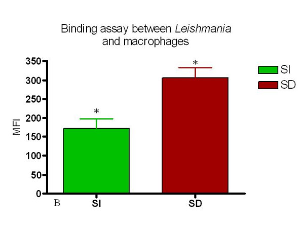
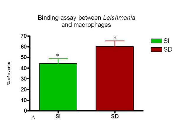
Results: Our results have shown that the interaction between Leishmania and macrophages can be measured by flow cytometry using the fluorescent dye CFSE to identify the Leishmania, and measuring simultaneously the expression of an important integrin involved in this interaction: the CD11b/CD18 (CR3 or Mac-1) beta2 integrin.

Conclusion: Flow cytometry offers rapid, reliable and sensitive measurements of single cell interactions with Leishmania in unstained or phenotypically defined cell populations following staining with one or more fluorochromes.


Figures

Figure 1
Figure 1
The use of CFSE stained- L. chagasi to evaluate parasite-canine macrophage interaction. L. chagasi promastigotes were stained with the intracellular dye CFSE and the efficiency of the staining procedure evaluated trough flow cytometry (A) and conventional fluorescence microscopy (B). CFSE-stained parasites were used for in vitro infection of peritoneal dog macrophages (X to Y ratio) and the parasite-macrophage interaction was evaluated through flow cytometry. Macrophages were selected based on forward- versus side-scatter parameters (C) and the frequency and intensity of CFSE+ macrophages was evaluated (D to F – MI Marker). At least 30,000 events were collected. (D) Non-infected macrophages. (E) Macrophages infected by CFSE-stained parasites in absence of C5 deficient sera. (F) Macrophages infected by CFSE-stained parasites in presence of C5 deficient sera. Data represents the mean ± SE of the frequency of infected macrophages and the CFSE fluorescence intensity of infected macrophages, obtained for one dog, representing identical experiments of four dogs.
Figure 2
Figure 2
Cells from peritoneal lavage adhered to coverslips. The image shows the predominance of macrophages (arrow) and neutrophils (arrow head). Giemsa stain – 400×.
Figure 3
Figure 3
Conventional Binding assay showing the interaction of macrophages from one dog with several Leishmaniae. Giemsa stain – 400×.
Figure 4
Figure 4
Conventional Binding assay showing the interaction of one macrophage with several Leishmaniae. Giemsa stain – 1000× optical microscope – 2× digitalized image.
Figure 7
Figure 7
Serum dependent (SD) conditions induce a higher association of Leishmania with macrophages as measured by both mean fluorescent intensity and percent macrophages associated with Leishmania. This figure shows the MFI of serum independent SI and SD. analysis. n = 4 animals. * = p < 0.05.
Figure 8
Figure 8
Percent of CFSE positive macrophages from SI and SD cultures. Canine macrophages were incubated for 1 hour with CFSE labeled Leishmania and analyzed using flow cytometry as described in Materials and Methods. n = 4 animals. * = p < 0.05.
Figure 5
Figure 5
Increased parasite/macrophage ratios leads to an increased intensity of macrophages associated with CFSE marked parasites. The intensity of fluorescence increases in accordace with the number of parasites per macrophage used. Macrophages and parasites were combined as described in Materials and Methods and analyzed for fluorescent intensity using flow cytometry. The data represent the mean flourescent intensity of posive macrophage populations from cultures at each ratio studied. These studies are representative of 4 experiments.
Figure 6
Figure 6
The levels of CD11b and CD18 fall dramatically following incubation with Leishmania under serum dependent (SD) culture conditions. Peritoneal macrophages were selected based on forward- versus side-scatter parameters as described in Materials and Methods and Figure 1 using flow cytometry. A) Isotype control with macrophages showing delineation of the M1 marker based on the negative population. B and C represent expression of CD18 (B) or CD11b (C) before incubation with Leishmania. D and E represent expression of CD18 (D) or CD11b (E) after association with Leishmania in a SD assay. In all cases the percent positive cells within the M1 marker are identified. This Figure is a representative diagram of 4 experiments with similar findings.
Figure 9
Figure 9
Percent peritoneal cells expressing CR3 as determined by expression of CD11b and CD18. Peritoneal cells from 4 dogs were stained ex vivo and analyzed using flow cytometry as described in Materials and Methods.
Figure 10
Figure 10
The expression of both CD11b and CD18 is dramatically reduced following association of peritoneal macrophages with Leishmania in both assays (SI or SD). Percent of CD11b expressing macrophages before and after incubation with Leishmania (SD assay).
Figure 11
Figure 11
The expression of both CD11b and CD18 is dramatically reduced following association of peritoneal macrophages with Leishmania in both assays (SI or SD). Percent of CD18 expressing macrophages before and after incubation with Leishmania (SD assay). Macrophages were incubated with or without Leishmania for one hour and stained with the appropriate antibody followed by analysis using flow cytometry as described in Materials and Methods. n = 4 animals, and * p < 0.05.

Similar articles

Cited by

References

    1. Lainson R, Shaw JJ. Epidemiology and ecology of leishmaniasis in Latin America. Nature. 1978;273:595. - PubMed
    1. Grimaldi G, Tesh RB, Mc Mathon-Pratt DA. A review of the geographic distribution and epidemiology of Leishmaniasis in the New World. Am J Trop Med Hyg. 1989;41:687. - PubMed
    1. Ashford RW. The Leishmaniasis as emerging and reemerging zoonoses. Int J Parasitol. 2000;30:1269. doi: 10.1016/S0020-7519(00)00136-3. - DOI - PubMed
    1. Sacks DL. Metacyclogenesis in Leishmania promastigotes. Exp Parasitol. 1989;69:100. doi: 10.1016/0014-4894(89)90176-8. - DOI - PubMed
    1. Schlein Y. Leishmania and Sandflies: Interactions In life cycle and transmission. Parasitol Today. 1993;9:255. doi: 10.1016/0169-4758(93)90070-V. - DOI - PubMed

Publication types