. 2005 Sep 1;106(5):1590-600.
doi: 10.1182/blood-2005-03-0860. Epub 2005 May 24.

Distinctive and indispensable roles of PU.1 in maintenance of hematopoietic stem cells and their differentiation

Affiliations

Distinctive and indispensable roles of PU.1 in maintenance of hematopoietic stem cells and their differentiation

Hiromi Iwasaki et al. Blood. .

Abstract

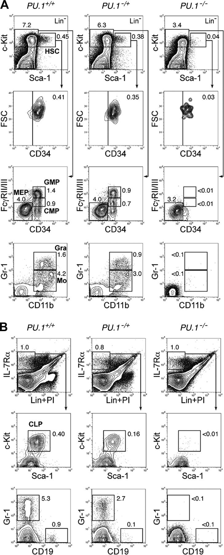
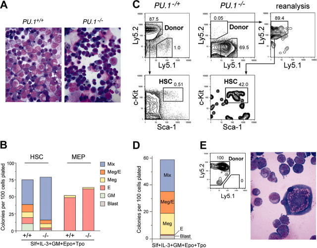
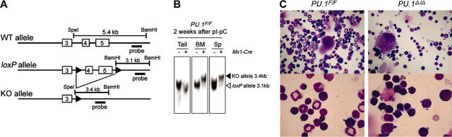
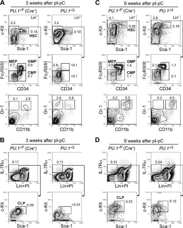
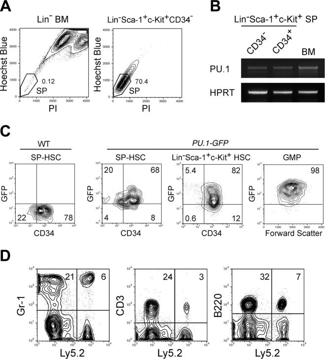
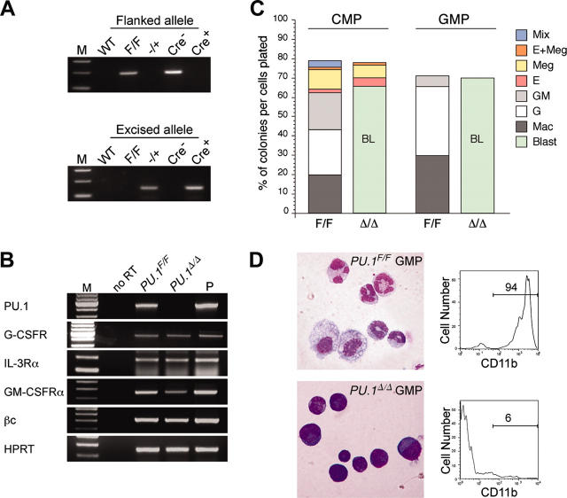
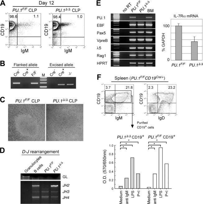
The PU.1 transcription factor is a key regulator of hematopoietic development, but its role at each hematopoietic stage remains unclear. In particular, the expression of PU.1 in hematopoietic stem cells (HSCs) could simply represent "priming" of genes related to downstream myelolymphoid lineages. By using a conditional PU.1 knock-out model, we here show that HSCs express PU.1, and its constitutive expression is necessary for maintenance of the HSC pool in the bone marrow. Bone marrow HSCs disrupted with PU.1 in situ could not maintain hematopoiesis and were outcompeted by normal HSCs. PU.1-deficient HSCs also failed to generate the earliest myeloid and lymphoid progenitors. PU.1 disruption in granulocyte/monocyte-committed progenitors blocked their maturation but not proliferation, resulting in myeloblast colony formation. PU.1 disruption in common lymphoid progenitors, however, did not prevent their B-cell maturation. In vivo disruption of PU.1 in mature B cells by the CD19-Cre locus did not affect B-cell maturation, and PU.1-deficient mature B cells displayed normal proliferation in response to mitogenic signals including the cross-linking of surface immunoglobulin M (IgM). Thus, PU.1 plays indispensable and distinct roles in hematopoietic development through supporting HSC self-renewal as well as commitment and maturation of myeloid and lymphoid lineages.


Figures

Figure 1.
Figure 1.
PU.1 knock-out fetal liver HSCs are arrested at the transition to the common myeloid and lymphoid progenitor stages. Multicolor FACS analysis was performed in E14.5 fetal livers from PU.1+/+, PU.1/+, and PU.1/ embryos. (A) The top panels demonstrate the Sca-1/c-Kit profile of Lin cells. PU.1/ fetal liver has a decreased number of LinSca-1+c-Kit+CD34+ HSCs. PU.1/ fetal liver lacks CMPs and GMPs as well as their progeny, mature monocytic (CD11b+/Gr-1lo) and granulocytic (CD11b+/Gr-1hi) populations. (B) PU.1+/+, PU.1/+, and PU.1/ fetal livers have almost equal numbers of LinIL-7Rα+ cells. PU.1/ fetal liver lacks LinIL-7Rα+Sca-1loc-Kitlo CLPs and CD19+ early B cells. In both analyses, PU.1/+ fetal liver has intermediate numbers of myeloid and lymphoid progenitors. Numbers in each panel represent percentages of the gated population in whole fetal liver cells. FSC indicates forward scatter; Gra, granulocytes; Mo, monocytes; and Lin, lineage.
Figure 2.
Figure 2.
Stem and progenitor activity of PU.1/ fetal liver cells. (A) Morphology of E14.5 fetal liver cells of PU.1+/+ and PU.1/ embryos (May-Giemsa staining, 600×). (B) The numbers of specific types of colonies derived from purified HSCs (left columns) and MEPs (right columns) from PU.1+/+ and PU.1/ fetal livers. Note that in PU.1/ cultures, there were no mature granulocytic and monocytic components that were replaced by immature myeloblastic cells. (C) Analysis of reconstitution activity of PU.1/ fetal liver HSCs. Twelve weeks after injection of high doses (1000 cells) of PU.1/ HSCs (Ly5.2+) into Ly5.1+ congenic lethally irradiated hosts, a minor population of donor-derived cells (Ly5.2+) of HSC phenotype (LinSca-1+c-Kit+) was detected. Control experiments using 100 PU.1/+ fetal liver HSCs (Ly5.2+) as a donor demonstrated that nearly all of the recipient bone marrow cells were of donor origin. (D) Colony assay of purified secondary PU.1/ HSCs. They displayed colony-forming activity almost equal to primary PU.1/ HSCs (B). (E) A mixed colony derived from single secondary PU.1/ HSC expressed the Ly5.2 donor maker, and contained erythroblasts and megakaryocytes and immature blastic myelomonocytic cells but not mature granulocytes or macrophages (May-Giemsa staining, 1000×).
Figure 3.
Figure 3.
Generation and characterization of conditional PU.1 knock-out mice. (A) Generation of a conditional targeted allele of the murine PU.1 gene. Cre recombination sites (loxP, black arrowheads) were inserted distal to the SpeI site in intron 3 and 435 base pair (bp) distal to the end of exon 5 as indicated. Shown are the predicted structures of the wild-type allele (WT), the targeted allele with the loxP sites (loxP), and the allele after Cre-mediated recombination, in which exons 4 and 5 have been deleted (KO). The relative location of the probe used in Southern blot analysis (a 1.3-kb BamHI/EcoRI fragment) is also shown. (B) Southern blot analysis of Mx1-Cre × PU.1F/F mice 2 weeks following pI-pC injection. BM indicates bone marrow. (C) Morphology of adult bone marrow cells in PU.1F/F (left) or Mx1-Cre × PU.1F/F (right) mice 3 weeks after injection of pI-pC (May-Giemsa staining; top panels: 400×; bottom panels: 1000×).
Figure 4.
Figure 4.
Conditional depletion of PU.1 in adult hematopoiesis. FACS analysis of Mx1-Cre × PU.1/F mice 3 (A-B) and 6 (C-D) weeks after pI-pC injection. Three weeks after the injection, HSCs were decreased up to 5-fold, and CMPs and GMPs disappeared (A). At this time point, the vast majority of bone marrow cells excised loxP-flanked PU.1 alleles (not shown). Significant recovery of these cells was observed at 6 weeks (C), when the excised allele of PU.1 locus was undetectable (not shown). CLPs were eliminated 3 weeks following disruption of PU.1 (B), while they recovered at the 6-week time point (D). Numbers in each panel represent percentages of the gated population in whole bone marrow cells. Summarized data including results of pI-pC injection into Mx1-Cre × PU.1F/F mice are shown in Table 1.
Figure 5.
Figure 5.
PU.1 is expressed in functional HSCs with long-term reconstituting potential. (A) Sorting gates for the SP fraction. The vast majority of LinSca-1+c-Kit+CD34 cells possessed the SP profile. (B) RT-PCR analysis of PU.1 mRNA in CD34+ or CD34 SP HSCs. HPRT indicates hypoxanthin phosphoribosyltransferase. (C) Expression of GFP in HSCs and GMPs purified from PU.1GFP/+ mice. The vast majority of LinSca-1+c-Kit+ HSCs and SP HSCs expressed PU.1-GFP irrespective of CD34 expression at the single-cell level. GMPs expressed GFP at a higher level compared with HSCs. (D) Ly5.2+ donor type Gr-1+, CD3+, and B220+ cells were successfully reconstituted in a mouse that received a transplant of a single CD34PU.1-GFP+ HSC. Multilineage reconstitution has been maintained for more than 20 weeks.
Figure 6.
Figure 6.
PU.1 disruption at the CMP or GMP stage. (A) PCR analysis of flanked PU.1 alleles in control and Cre-transduced GMPs. The flanked PU.1 allele became undetectable after the Cre transduction into PU.1F/F GMPs. M indicates the molecular weight marker. (B) RT-PCR analyses of cytokine receptors in PU.1Δ/Δ CMPs. M indicates the molecular weight marker; P, the positive control. (C) Myeloid colony assays of single sorted PU.1F/F and PU.1Δ/Δ CMPs/GMPs in the presence of SCF, GM-CSF, IL-3, Epo, and Tpo. PU.1Δ/Δ CMPs and GMPs could not form mature M, G, or GM colonies, but formed myeloblast colonies. BL indicates blast. (D) PU.1Δ/Δ GMPs formed colonies composed mainly of immature myeloid cells (May-Giemsa staining, 600×), which expressed only low levels of CD11b.
Figure 7.
Figure 7.
PU.1 disruption at the CLP stage. (A) B-cell differentiation assays of PU.1F/F and PU.1Δ/Δ CLPs in the presence of IL-7. PU.1F/F and PU.1Δ/Δ CLPs gave rise to almost equal numbers of CD19+IgM+ mature B cells after 12 days of culture on OP9 cells in the presence of IL-7. (B) PU.1Δ/Δ CLP-derived B-cell progeny completely excised floxed alleles. (C) PU.1F/F and PU.1Δ/Δ CLPs gave rise to similar sizes of B-cell colonies in response to IL-7 after 12 days in culture. (D) Both PU.1Δ/Δ and PU.1F/F CLP-derived B cells rearranged their IgH gene. (E) RT-PCR analyses of B-cell–related genes in PU.1F/F and PU.1Δ/Δ B cells (left). IL-7Rα transcripts were quantitated by a real-time PCR analysis (right). Error bars indicate SD. (F) Analysis of spleen B cells developed in PU.1F/FCD19Cre/+ mice. PU.1Δ/Δ CD19+ B cells expressed normal levels of IgM and IgD (upper panels). Purified PU.1Δ/Δ CD19+ B cells from PU.1F/FCD19Cre/+ mice displayed normal proliferative response to mitogenic agents including anti-IgM antibodies, LPS, and PMA plus ionomycin (P+I) determined by an MTT assay (bottom). *P < .05.

References

    1. Shivdasani RA, Orkin SH. The transcriptional control of hematopoiesis. Blood. 1996;87: 4025-4039. - PubMed
    1. Tenen DG, Hromas R, Licht JD, Zhang DE. Transcription factors, normal myeloid development, and leukemia. Blood. 1997;90: 489-519. - PubMed
    1. Klemsz MJ, McKercher SR, Celada A, Van Beveren C, Maki RA. The macrophage and B cell-specific transcription factor PU.1 is related to the ets oncogene. Cell. 1990;61: 113-124. - PubMed
    1. Akashi K, Traver D, Miyamoto T, Weissman IL. A clonogenic common myeloid progenitor that gives rise to all myeloid lineages. Nature. 2000;404: 193-197. - PubMed
    1. Miyamoto T, Iwasaki H, Reizis B, et al. Myeloid or lymphoid promiscuity as a critical step in hematopoietic lineage commitment. Dev Cell. 2002;3: 137-147. - PubMed

Publication types