. 2005 Jun;46(6):2156-67.
doi: 10.1167/iovs.04-1427.

Cone-like morphological, molecular, and electrophysiological features of the photoreceptors of the Nrl knockout mouse

Affiliations

Cone-like morphological, molecular, and electrophysiological features of the photoreceptors of the Nrl knockout mouse

Lauren L Daniele et al. Invest Ophthalmol Vis Sci. 2005 Jun.

Abstract

Purpose: To test the hypothesis that Nrl(-)(/)(-) photoreceptors are cones, by comparing them with WT rods and cones using morphological, molecular, histochemical, and electrophysiological criteria.

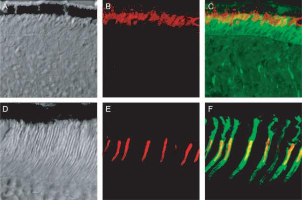
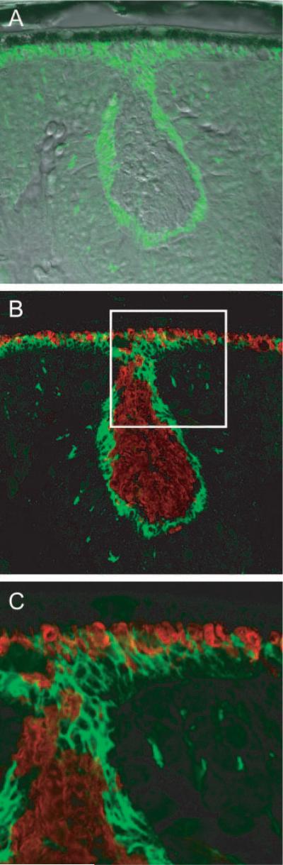
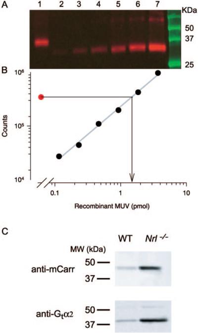
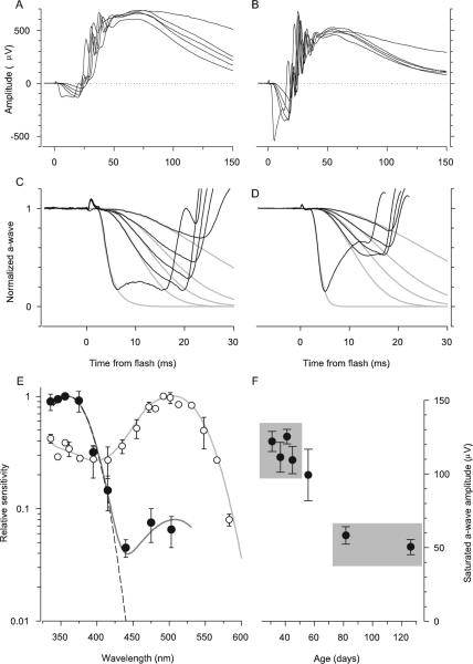
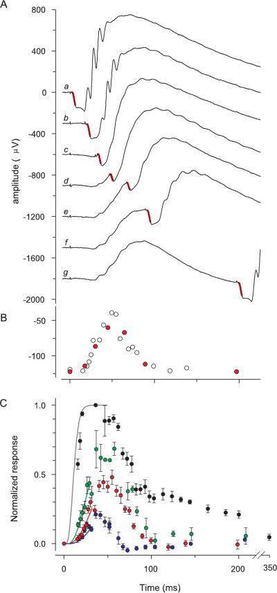
Methods: The photoreceptor layer of fixed retinal tissue of 4- to 6-week-old mice was examined in plastic sections by electron microscopy, and by confocal microscopy in frozen sections immunolabeled for the mouse UV-cone pigment and colabeled with PNA. Quantitative immunoblot analysis was used to determine the levels of expression of key cone-specific proteins. Single- and paired-flash methods were used to extract the spectral sensitivity, kinetics, and amplification of the a-wave of the ERG.

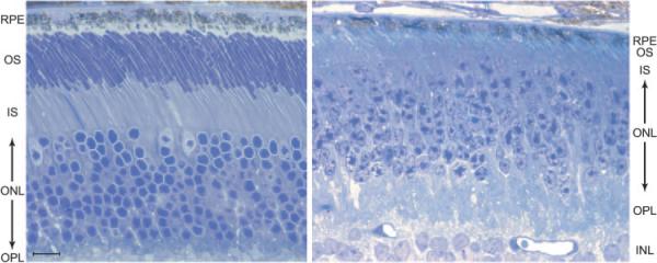
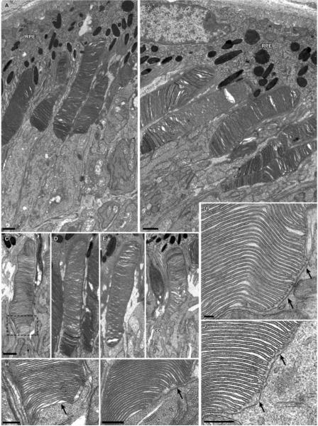
Results: Outer segments of Nrl(-/-) photoreceptors ( approximately 7 mum) are shorter than those of wild-type (WT) rods ( approximately 25 mum) and cones ( approximately 15 mum); but, like WT cones, they have 25 or more basal discs open to the extracellular space, extracellular matrix sheaths stained by PNA, chromatin "clumping" in their nuclei, and mitochondria two times shorter than rods. Nrl(-/-) photoreceptors express the mouse UV cone pigment, cone transducin, and cone arrestin in amounts expected, given the relative size and density of cones in the two retinas. The ERG a-wave was used to assay the properties of the photocurrent response. The sensitivity of the Nrl(-/-) a-wave is at its maximum at 360 nm, with a secondary mode at 510 nm having approximately one-tenth the maximum sensitivity. These wavelengths are the lambda(max) of the two mouse cone pigments. The time to peak of the dim-flash photocurrent response was approximately 50 ms, more than two times faster than that of rods.

Conclusions: Many morphological, molecular, and electrophysiological features of the Nrl(-/-) photoreceptors are cone-like, and strongly distinguish these cells from rods. This retina provides a model for the investigation of cone function and cone-specific genetic disease.


Figures

Figure 1
Figure 1
Light micrograph of a semithin section of the RPE and photoreceptor layers of the retina of a 5-week-old Nrl−/− mouse (right) and age-matched WT mouse (left). Note the three cone nuclei (pale-staining nucleoplasm) in the outermost layer of the ONL of the WT retina. RPE, retinal pigment epithelium cell layer; OS, outer segment layer; IS, inner segment layer; ONL, outer nuclear layer; OPL, outer plexiform; INL, inner nuclear layer. Bar, 10 μm.
Figure 2
Figure 2
Electron micrographs of photoreceptor cells from Nrl−/− retinas. (A, B) Groups of photoreceptor cells and adjacent RPE. (C–F) Examples of individual photoreceptor outer segments. Although less organized than normal outer segments, the disc membranes form a well-defined stack. Outlined area in (C) is shown at higher magnification in (H). (G–J) Higher magnification of the basal region of the outer segments from several photoreceptor cells. Arrows: inner segment membrane apposes the basal discs, which are open to the extracellular space; the region of open discs extends much further distally than observed for normal rod outer segments (cf. Ref. 28). RPE, retinal pigmented epithelium; CC, connecting cilium. Scale bars: (A–F) 1 μm; (G–J) 300 nm.
Figure 3
Figure 3
(A–C) Images of a frozen section of Nrl−/− retina taken with differential interference contrast optics (A), with fluorescent immuno-staining of MUV, the mouse ultraviolet pigment (B, red), and with overlaid fluorescent immunostaining of MUV and PNA binding (green) (C). (D–F) Images of a frozen section of a WT retina stained and presented in the same format as in (A–C). Images were made with a confocal microscope and represent 50 × 50-μm regions of the two retinas.
Figure 4
Figure 4
Confocal images of a rosette in the ONL of the Nrl−/− retina. (A) Differential interference contrast image with overlaid image taken of PNA labeling (green). (B) Same section as in (A), but merging immunostaining of MUV (red) and PNA labeling. MUV is seen to fill the center portion of the rosette; the white square is 50 × 50 μm. (C) Magnified image of the portion of (B) highlighted by the white square, showing details of a portion of the rosette. Note how the PNA labeling of the border of the rosette merges with the PNA-stained inner segment layer of the cones that are in contact with the RPE.
Figure 5
Figure 5
Immunoblot analysis of cone phototransduction cascade molecules in the Nrl−/− retina. (A) Immunoblot of the mouse UV-cone pigment (MUV) of a 4-week-old Nrl−/− mouse: lane 1: 1% of the lysate of two Nrl−/− eyes; lanes 2 to 7: twofold incremented amounts of recombinant MUV; the latter runs at a slightly lower molecular mass because of mutations engineered to allow it to be purified with a commercial antibody. (B) Plot of the blot densities in (A): each point (●) corresponds to the density of the MUV immunolabeling in the blot lane immediately above it in (A). The symbol corresponding to lane 1 yields the estimate of the MUV mass loaded from the Nrl−/− eye, 1.6 picomoles (arrow projecting to abscissa). Because 1% of the lysate of two eyes was loaded in lane 1, the MUV per eye is thus estimated to be 80 picomoles. (C) Blots comparing extracts of Nrl−/− and WT mouse eyes for cone arrestin (mCarr) and the α-subunit of cone transducin (Gtα2): 25 μg of protein from eyes of animals of each genotype was loaded into adjacent lanes of the gel and immunolabeled. The blot densities of regions circumscribing the immunolabel were quantified, and the ratio of the densities for the blots of the WT and Nrl−/− lanes were determined. The ratios were 14:1 for the mCarr comparison and 11:1 for the Gtα2 comparison in the blots illustrated. Mean ratios (± SEM) were 12.3 ± 1.1 for mCarr (two blots, 11 comparisons of proteins from five Nrl−/− and two WT mice), and 14.1 ± 2.4 for Gtα2 (three blots, 21 comparisons).
Figure 6
Figure 6
Comparison of the properties of the ERG a-wave responses of Nrl−/− and WT mice. (A) ERGs of an Nrl−/− mouse obtained in response to a series of 360-nm flashes that produced a-waves. The flash intensities were 7,400, 21,000, 36,400, 68,000 in photons/μm2 at the cornea. The most intense flash was an unattenuated white flash and saturated the a-wave amplitude. (B) ERGs of a WT mouse obtained in response to the same series of flashes as was used in the experiment in (A). The a-wave components of the responses of the Nrl−/− mouse in (A) have been extracted and normalized and fitted with a model of the activation phase of phototransduction (C),, modified to incorporate the membrane time constant, which was set to 2 ms. The amplification constant obtained from fitting the model is A = 3.6 s−2 (cf. equation 1). (D) The a-wave components of the responses of the WT mouse (B) have been normalized and analyzed with the model, with a membrane time constant of 1 ms. The amplification constant of the same mouse obtained from fitting the cascade model to the a-wave responses to 500-nm flashes (data not shown) is A = 10 s−2; the theory traces in (D) were obtained with this value of A, and a spectral sensitivity factor at 360 nm of S360 = 0.3. (E) Spectral sensitivities of the a-wave of Nrl−/− (●) and WT mice (○). Results such as those shown in (C) and (D) were analyzed as described in Lyubarsky et al. to extract the spectral sensitivities. Dark gray curve through the Nrl−/− data was derived by combining a pigment template having λmax = 360 nm and unit sensitivity with a second template with λmax = 508 nm and maximum sensitivity of 0.08; dashed curve: 360-nm template alone. The light gray curve through the rod data (○) is a 500-nm template above 470 nm and a fifth-order spline below 470 nm. The rod data and template curves are taken from Lyubarsky et al. (F) The saturating amplitude amax of the a-wave of Nrl−/− mice as a function of age, derived from experiments such as illustrated in (A). Each point is the mean ± SEM of a group of 6 to 10 mice (except the initial point, for which n = 5) and is plotted at the mean age of the group. For each mouse the responses from both eyes to at least 5 (but up to 20) saturating flashes were averaged to estimated amax. Gray rectangles identify two groups of mice: average age 39 days (top left; n = 33); average age 104 days (bottom right; n = 13). The difference in amax between these two groups was significant at P < 10−9 (two-sample t-test, df = 35).
Figure 7
Figure 7
Kinetics of the photocurrent response of the Nrl−/− mouse derived with the paired-flash ERG method. (A) Series of traces (b–g) from experiment in which a single 360-nm test flash was delivered at t = 0 followed by an intense probe flash that saturated the a-wave amplitude. Trace a: response to the probe alone, delivered at t = 0; trace g: an almost complete response to the test flash. The portion of the traces highlighted in red is the a-wave component of the response to the probe flash, and gives a measure of the residual photoreceptor circulating current present at various times after the test flash (i.e., that not suppressed by the test flash). Each trace is the average of 7 repetitions of a series of 24 combinations of the test and probe flashes. (B) Amplitudes of the responses to the probe flash in the experiment of (A), plotted as a function of the time of its delivery; red symbols: amplitudes derived from the seven traces illustrated in (A); open symbols: other data collected at other test–probe intervals in the same experiment. (C) Results from paired-flash experiments involving 360-nm test flashes of different strengths. The intensities (in photons per square micrometer at the cornea) and number of animals (n) whose data were averaged were 3,900 (11, blue symbols), 7,300 (6, red symbols), 23,300 (6, green symbols), 40,000 (3, black symbols). The smooth curves are derived from the activation model of photo-transduction with A = 7.5 s−2. The curves are color coded in correspondence to the respective sets of data.

References

    1. Swaroop A, Xu JZ, Pawar H, et al. A conserved retina-specific gene encodes a basic motif/leucine zipper domain. Proc Natl Acad Sci USA. 1992;89:266–270. - PMC - PubMed
    1. Swain PK, Hicks D, Mears AJ, et al. Multiple phosphorylated iso-forms of NRL are expressed in rod photoreceptors. J Biol Chem. 2001;276:36824–36830. - PubMed
    1. Yoshida S, Mears AJ, Friedman JS, et al. Expression profiling of the developing and mature Nrl−/− mouse retina: identification of retinal disease candidates and transcriptional regulatory targets of Nrl. Hum Mol Genet. 2004;13:1487–1503. - PubMed
    1. Bessant DA, Payne AM, Mitton KP, et al. A mutation in NRL is associated with autosomal dominant retinitis pigmentosa. Nat Genet. 1999;21:355–356. - PubMed
    1. Mears AJ, Kondo M, Swain PK, et al. Nrl is required for rod photoreceptor development. Nat Genet. 2001;29:447–452. - PubMed

Publication types

MeSH terms