. 2005 May 24;33(9):e86.
doi: 10.1093/nar/gni085.

A versatile reducible polycation-based system for efficient delivery of a broad range of nucleic acids

Affiliations

A versatile reducible polycation-based system for efficient delivery of a broad range of nucleic acids

Martin L Read et al. Nucleic Acids Res. .

Abstract

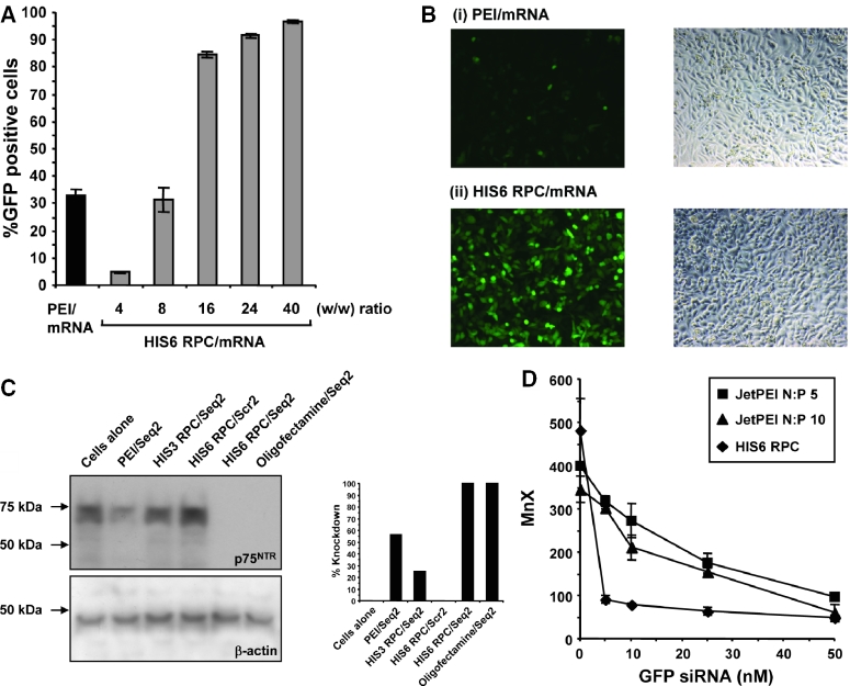
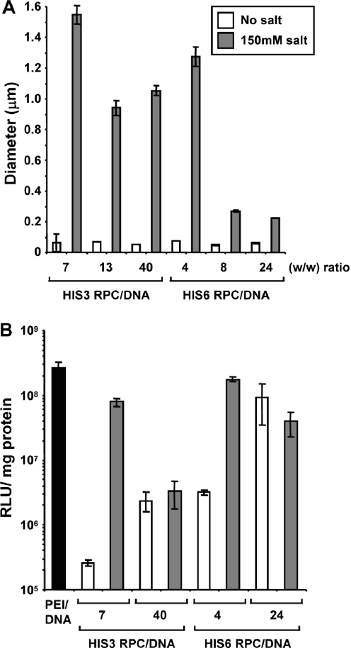
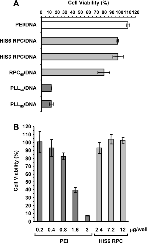
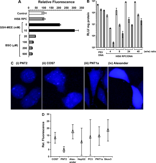
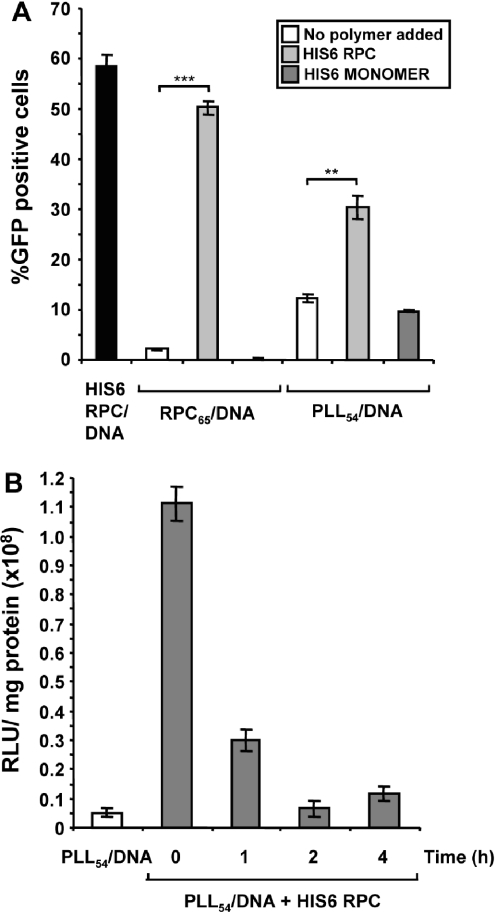
Synthetic vectors based on reducible polycations consisting of histidine and polylysine residues (HIS RPCs) were evaluated for their ability to deliver nucleic acids. Initial experiments showed that RPC-based vectors with at least 70% histidine content mediated efficient levels of gene transfer without requirement for the endosomolytic agent chloroquine. Significant gene transfer was observed in a range of cell types achieving up to a 5-fold increase in the percentage of transfected cells compared to 25 kDa PEI, a gold standard synthetic vector. In contrast to 25 kDa PEI, HIS RPCs also mediated efficient transfer of other nucleic acids, including mRNA encoding green fluorescent protein in PC-3 cells and siRNA directed against the neurotrophin receptor p75(NTR) in post-mitotic cultures of rat dorsal root ganglion cell neurons. Experiments to elevate intracellular glutathione and linear profiling of cell images captured by multiphoton fluorescent microscopy highlighted that parameters such as the molecular weight and rate of cleavage of HIS RPCs were important factors in determining transfection activity. Altogether, these results demonstrate that HIS RPCs represent a novel and versatile type of vector that can be used for efficient cytoplasmic delivery of a broad range of nucleic acids. This should enable different or a combination of therapeutic strategies to be evaluated using a single type of polycation-based vector.


Figures

Figure 1
Figure 1
Characterization of HIS RPCs and polyplex formation. (A) Mwt growth of oxidative polymerization of HIS6 RPC in DMSO over time. Dialysis of HIS RPCs was performed in Centricon filters (10 000 mwt cut off) with a final volume of 400–500 μl. (B) pH titration curves of histidine residues of HIS6 RPC measured by NMR spectroscopy. The pH titration was achieved using 10 μM HIS6 RPC dissolved in deuterated water and basified using deuterated sodium hydroxide. The chemical shift for the H5 and H6 protons in the imidazole ring were plotted (relative positions of H5 and H6 protons are shown in inset). (C) EtBr exclusion assay was performed as previously described (21) with HIS6 RPC (filled squares) and HIS3 RPC (filled diamonds). Results are shown as mean and SD values from at least three samples. (D) Agarose gel electrophoresis of pCMVLuc1 complexed with HIS6 RPC at the indicated (w/w) ratio. The control lane (-) contains DNA alone. The relative locations of wells and free DNA are indicated.
Figure 2
Figure 2
Reduction destabilizes HIS6 RPC/DNA polyplexes. To determine the ability of polyplexes to be activated by reduction HIS6 RPC/DNA polyplexes formed at (w/w) ratio of 4 were incubated with 25 mM DTT for 1 h. NaCl was added at a range of concentrations (0–1.0 M) and the amount of free DNA assessed by gel electrophoresis. ImageQuant™ software was used to quantify the amount of DNA released (Rel. = relaxed DNA; S.C. = supercoiled DNA).
Figure 3
Figure 3
Histidine-rich RPCs facilitate chloroquine-independent gene transfer. PC-3 cells were transfected with 0.5 μg pCMVLuc condensed by either (A) HIS 3 RPC or (B) HIS6 RPC at the indicated (w/w) ratio. For control transfections 25 kDa PEI/DNA polyplexes were formed at N:P 10. Cells were incubated with polyplexes for 4 h in the absence (white bars) or presence (dark gray bars) of 100 μM chloroquine and the media was discarded and replaced with fresh media containing 10% FCS. Luciferase activity was measured after 24 h. Results are shown as mean and SD values from at least three samples.
Figure 4
Figure 4
Enhanced frequency of gene transfer with histidine-rich RPCs. (A) PC-3 cells were transfected with 0.5 μg pEGFPN1 condensed by either HIS3 RPC, HIS6 RPC, HIS6 monomer or RPC65 at the indicated (w/w) ratio, and by 25 kDa PEI at N:P 10 (P = 0.0005). (B) Fluorescent images of PC-3 cells expressing GFP following transfection with (i) nothing or 0.5 μg pEGFPN1 condensed with (ii) 25 kDa PEI at N:P 10 or (iii) HIS6 RPC at (w/w) ratio of 40. Phase-contrast images of transfected PC-3 cells are shown in the corresponding right-hand column. (C) A range of cell types were transfected as indicated with 0.5 μg pEGFPN1 condensed by 25 kDa PEI at N:P 10 (white bars), HIS6 RPC at (w/w) ratio of 40 (light grey bars) or HIS3 RPC at (w/w) ratio of 66 (dark grey bars). GFP expression was measured after 24 h by flow cytometry analysis. Results are shown as mean and SD values from at least three samples. Significant differences between HIS6 RPC/DNA and 25 kDa PEI/DNA transfected cells are indicated (***, P ≤ 0.0001; **, P ≤ 0.001; *, P ≤ 0.01).
Figure 5
Figure 5
Histidine-rich RPCs mediate efficient delivery of mRNA and siRNA. (A) PC-3 cells were transfected with 0.5 μg cap-GFP-A64 mRNA condensed either by PEI at N:P 10 (black bar) or HIS6 RPC at the indicated (w/w) ratio (dark grey bars). (B) Fluorescent images of PC-3 cells expressing GFP following transfection with 0.5 μg cap-GFP-A64 mRNA condensed with (i) 25 kDa PEI at N:P 10 or (ii) HIS6 RPC at (w/w) ratio of 40. Phase-contrast images of transfected PC-3 cells are shown in the corresponding right-hand column. (C) Western blot analysis of p75NTR (upper panel) and β-actin (lower panel) using DRG culture lysates following transfection with siRNA directed against p75NTR (Seq2) or a scrambled sequence (Scr2) and condensed either by 25 kDa PEI at N:P 10, HIS3 RPC at (w/w) ratio of 66, HIS6 RPC at (w/w) ratio of 40 or Oligofectamine at (w/w) ratio of 3.5 as indicated. The relative% knockdowns in p75NTR levels are shown in the bar graph on the right-hand side. (D) PC-3 cells were co-transfected with 0.5 μg pEGFPN1 and GFP-22 siRNA at the indicated dose condensed either by JetPEI at N:P 5 (squares), JetPEI at N:P 10 (triangles) or HIS6 RPC at (w/w) ratio of 24 (diamonds). In panels (A) and (D), flow cytometry analysis was used to determine %GFP positive cells and mean fluorescence per cell (MnX) after 24 h. Results are shown as mean and SD values from at least three samples.
Figure 6
Figure 6
Effect of salt-induced aggregation of particles on transfection activity. (A) Photon correlation spectroscopy was used to assess changes in the diameter of polyplexes formed between DNA condensed with either HIS3 RPC or HIS6 RPC at the indicated (w/w) ratio in the absence (white bars) or presence (dark grey bars) of 150 mM NaCl for 2.5 h. (B) PC-3 cells were transfected with 0.5 μg pCMVLuc1 condensed by either PEI at N:P 10 (black bar), HIS3 RPC or HIS6 RPC at the indicated (w/w) ratio. Polyplexes were incubated in the absence (white bars) or presence of 150 mM NaCl for 30 min (dark grey bars) prior to addition to cells. Luciferase activity was measured after 24 h. Results are shown as mean and SD values from at least three measurements.
Figure 7
Figure 7
Cellular toxicity of PEI and histidine-rich RPCs. (A) PC-3 cells were transfected with 0.5 μg of pCMVLuc1 condensed either by 25 kDa PEI at N:P 10 or HIS6 RPC, HIS3 RPC, RPC65, PLL54 and PLL85 at (w/w) ratio of 40. The cells were incubated with polyplexes for 4 h in serum-free media and the media discarded and replaced with fresh media containing 10% FCS. The MTS assay was used to assess the viability of transfected PC-3 cells after 24 h. (B) PC-3 cells were incubated in the presence of 25 kDa PEI (dark grey bars) or HIS6 RPC (light grey bars) at the indicated dose for 4 h in serum-free media. The media was discarded and replaced with fresh media containing 10% FCS and the MTS assay used to assess cellular viability after 24 h. Results are shown as mean and SD values from at least three samples.
Figure 8
Figure 8
Effect of intracellular GSH levels on transfection activity. (A) PC-3 cells were incubated in the presence of GSH–MEE for 3 h (black bars) or BSO for 24 h (dark grey bars) at the indicated concentrations and GSH levels determined as described by Sebastia et al. (22). For control experiments, cells were incubated on their own (white bars) or in the presence of HIS6 RPC (light grey bar). (B) PC-3 cells were incubated for 3 h in serum-free media containing 0 mM (white bars), 5 mM (light grey bars) or 10 mM GSH–MEE (dark grey bars) prior to transfection with 0.5 μg pCMV-Luc1 condensed with either 25 kDa PEI at N:P 10 or HIS6 RPC at the indicated (w/w) ratio. Luciferase activity was measured after 24 h. Results are shown as mean and SD values from at least three samples. (C) Microscopic comparison of GSH distribution in isolated (i) PNT2, (ii) COS7, (iii) PNT1a and (iv) Alexander cells based on mBCl fluorescence. (D) Relative fluorescence of mBCl-treated cells obtained by linear profiling from at least ten stacks in the indicated cell type. The empty triangle represents the mean fluorescence, whereas vertical bars represent the maximum and minimum levels of fluorescence. Background fluorescence was subtracted from the mean value.
Figure 9
Figure 9
Free HIS6 RPC enhances PLL54- and RPC65-mediated gene transfer. (A) PC-3 cells were transfected with 0.5 μg pEFGPN1 condensed with HIS6 RPC at (w/w) ratio of 40, RPC65 at (w/w) ratio of 2 or PLL54 at (w/w) ratio of 2. In these experiments, polyplexes were added to cells on their own (white bars) or by prior addition of free HIS6 RPC (light grey bars) or HIS6 monomer (dark grey bars). (B) Same as in (A) except that free HIS6 RPC was added to PC-3 cells at the indicated number of hours prior to transfection with 0.5 μg pCMV-Luc1 condensed with PLL54 at (w/w) ratio of 2. Luciferase activity was measured after 24 h. Results are shown as mean and SD values from at least three samples. Significant differences between RPC/DNA and PLL/DNA transfected cells in the absence or presence of HIS6 RPC are indicated (***, P ≤ 0.0001; **, P = 0.0002).
Figure 10
Figure 10
Proposed mechanism for HIS RPC-mediated delivery of nucleic acids. HIS RPC/DNA polyplexes are first internalized by endocytosis (1) and mediate escape from endocytic vesicles into the cytoplasm (2) thus avoiding degradation in lysosomal compartments (3). In the case of HIS3 RPC-based vectors, endosomal escape can be triggered by the buffering agent chloroquine (4). Intracellular reduction of HIS RPC-based polyplexes enables efficient cytoplasmic release (5) enabling transcription (6), translation (7) or RNA interference (8) to proceed depending on the nucleic acid payload.

References

    1. Dorsett Y., Tuschl T. siRNAs: applications in functional genomics and potential as therapeutics. Nature Rev. Drug. Discov. 2004;3:318–329. - PubMed
    1. Buckley R.H. Gene therapy for SCID—a complication after remarkable progress. Lancet. 2002;360:1185–1186. - PubMed
    1. European Society of Gene Therapy. French gene therapy group reports on the adverse event in a clinical trial of gene therapy for X-linked severe combined immune deficiency (X-SCID) J. Gene Med. 2003;5:82–84. - PubMed
    1. Boussif O., Lezoualc'h F., Zanta M.A., Mergny M.D., Scherman D., Demeneix B., Behr J.P. A versatile vector for gene and oligonucleotide transfer into cells in culture and in vivo: polyethylenimine. Proc. Natl Acad. Sci. USA. 1995;92:7297–7301. - PMC - PubMed
    1. Wagner E., Zenke M., Cotten M., Beug H., Birnstiel M.L. Transferrin-polycation conjugates as carriers for DNA uptake into cells. Proc. Natl Acad. Sci. USA. 1990;87:3410–3414. - PMC - PubMed

Publication types