

DNA damage response and Ku80 function in the vertebrate embryo
- PMID: 15914672
- PMCID: PMC1140083
- DOI: 10.1093/nar/gki613
DNA damage response and Ku80 function in the vertebrate embryo
Abstract
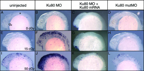
Cellular responses to DNA damage reflect the dynamic integration of cell cycle control, cell-cell interactions and tissue-specific patterns of gene regulation that occurs in vivo but is not recapitulated in cell culture models. Here we describe use of the zebrafish embryo as a model system to identify determinants of the in vivo response to ionizing radiation-induced DNA damage. To demonstrate the utility of the model we cloned and characterized the embryonic function of the XRCC5 gene, which encodes Ku80, an essential component of the nonhomologous end joining pathway of DNA repair. After the onset of zygotic transcription, Ku80 mRNA accumulates in a tissue-specific pattern, which includes proliferative zones of the retina and central nervous system. In the absence of genotoxic stress, zebrafish embryos with reduced Ku80 function develop normally. However, low dose irradiation of these embryos during gastrulation leads to marked apoptosis throughout the developing central nervous system. Apoptosis is p53 dependent, indicating that it is a downstream consequence of unrepaired DNA damage. Results suggest that nonhomologous end joining components mediate DNA repair to promote survival of irradiated cells during embryogenesis.
Figures


Similar articles
-
Expression of the Ku70 subunit (XRCC6) and protection from low dose ionizing radiation during zebrafish embryogenesis.Neurosci Lett. 2007 Jul 11;422(2):97-102. doi: 10.1016/j.neulet.2007.05.045. Epub 2007 Jun 2. Neurosci Lett. 2007. PMID: 17630212 Free PMC article.
-
The non-homologous end-joining pathway is not involved in the radiosensitization of mammalian cells by heat shock.J Cell Physiol. 2003 Sep;196(3):557-64. doi: 10.1002/jcp.10334. J Cell Physiol. 2003. PMID: 12891712
-
Quantification of ionizing radiation-induced cell death in situ in a vertebrate embryo.Radiat Res. 2007 Aug;168(2):149-57. doi: 10.1667/RR0803.1. Radiat Res. 2007. PMID: 17638406
-
The Ku heterodimer: function in DNA repair and beyond.Mutat Res Rev Mutat Res. 2015 Jan-Mar;763:15-29. doi: 10.1016/j.mrrev.2014.06.002. Epub 2014 Jul 4. Mutat Res Rev Mutat Res. 2015. PMID: 25795113 Review.
-
[Role of Ku protein in normal and cancer cells].Ukr Biokhim Zh (1999). 2006 Sep-Oct;78(5):5-15. Ukr Biokhim Zh (1999). 2006. PMID: 17290777 Review. Ukrainian.
Cited by
-
Zebrafish Ubc13 is required for Lys63-linked polyubiquitination and DNA damage tolerance.Mol Cell Biochem. 2010 Oct;343(1-2):173-82. doi: 10.1007/s11010-010-0511-9. Epub 2010 Jun 16. Mol Cell Biochem. 2010. PMID: 20556485
-
Effect of Photon Hormesis on Dose Responses to Alpha Particles in Zebrafish Embryos.Int J Mol Sci. 2017 Feb 11;18(2):385. doi: 10.3390/ijms18020385. Int J Mol Sci. 2017. PMID: 28208665 Free PMC article.
-
Effects of low-dose ionizing radiation and menadione, an inducer of oxidative stress, alone and in combination in a vertebrate embryo model.Radiat Res. 2012 Nov;178(5):499-503. doi: 10.1667/RR3042.2. Epub 2012 Oct 23. Radiat Res. 2012. PMID: 23092554 Free PMC article.
-
Banp regulates DNA damage response and chromosome segregation during the cell cycle in zebrafish retina.Elife. 2022 Aug 9;11:e74611. doi: 10.7554/eLife.74611. Elife. 2022. PMID: 35942692 Free PMC article.
-
Cellular responses to ionizing radiation change quickly over time during early development in zebrafish.Cell Biol Int. 2019 May;43(5):516-527. doi: 10.1002/cbin.11117. Epub 2019 Mar 12. Cell Biol Int. 2019. PMID: 30791195 Free PMC article.
References
-
- Ward J.F. Radiation mutagenesis: the initial DNA lesions responsible. Radiat. Res. 1995;142:362–368. - PubMed
-
- Collis S.J., Deweese T.L., Jeggo P.A., Parker A.R. The life and death of DNA-PK. Oncogene. 2005;24:949–961. - PubMed
-
- Zhivotovsky B., Kroemer G. Apoptosis and genomic instability. Nat. Rev. Mol. Cell Biol. 2004;5:752–762. - PubMed
-
- Streffer C., Shore R., Konermann G., Meadows A., Uma Devi P., Preston J., Holm L.E., Stather J., Mabuchi K., Withers H.R. Biological effects after prenatal irradiation (embryo and fetus). A report of the International Commission on Radiological Protection. Ann. ICRP. 2003;33:5–206. - PubMed
Publication types
MeSH terms
Substances
Associated data
- Actions
LinkOut - more resources
Full Text Sources
Molecular Biology Databases
Research Materials
Miscellaneous
