. 2005 Jul 22;280(29):27458-65.
doi: 10.1074/jbc.M502319200. Epub 2005 May 24.

Uncovering the molecular mode of action of the antimalarial drug atovaquone using a bacterial system

Affiliations

Uncovering the molecular mode of action of the antimalarial drug atovaquone using a bacterial system

Michael W Mather et al. J Biol Chem. .

Abstract

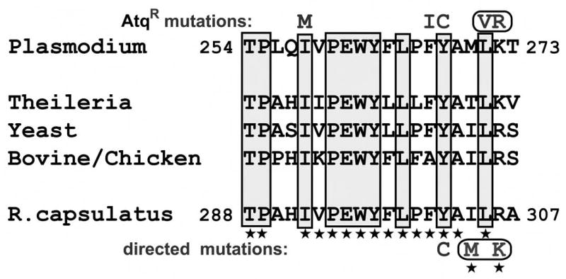
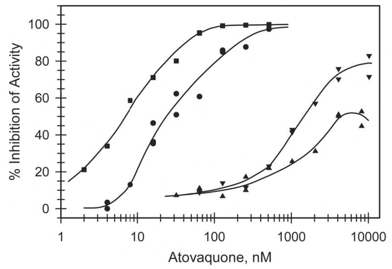
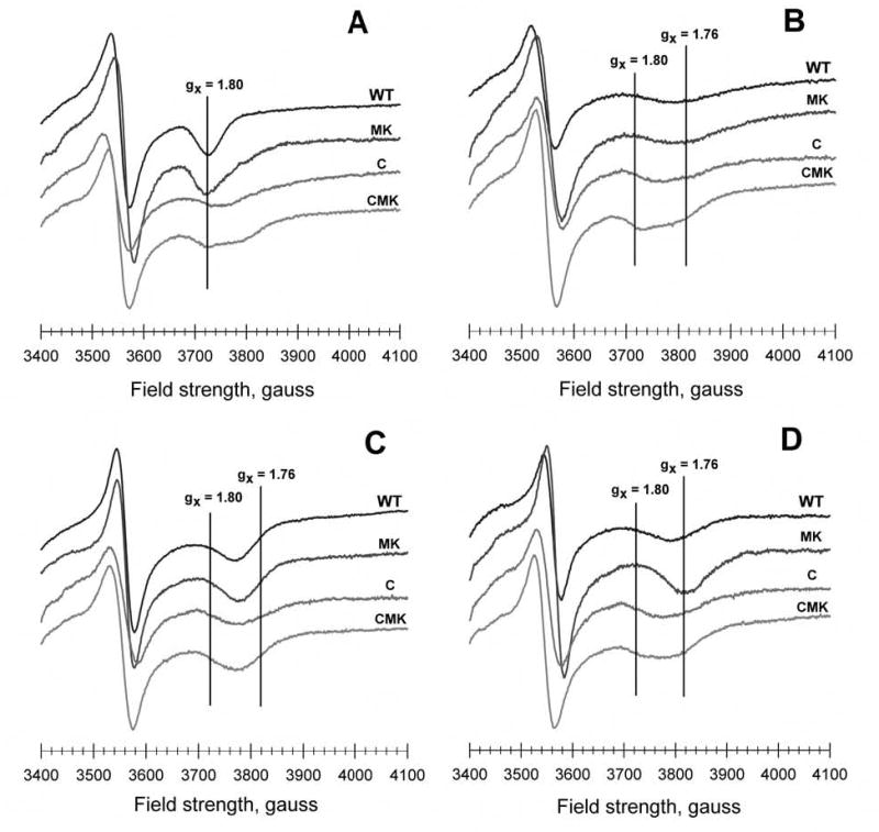
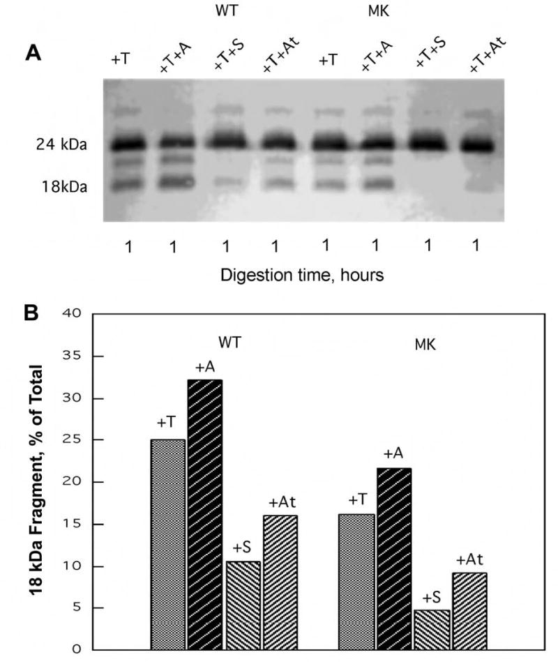
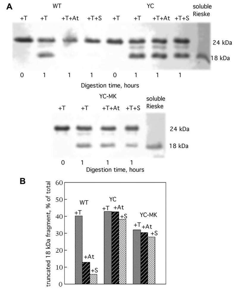
Atovaquone is an antiparasitic drug that selectively inhibits electron transport through the parasite mitochondrial cytochrome bc1 complex and collapses the mitochondrial membrane potential at concentrations far lower than those at which the mammalian system is affected. Because this molecule represents a new class of antimicrobial agents, we seek a deeper understanding of its mode of action. To that end, we employed site-directed mutagenesis of a bacterial cytochrome b, combined with biophysical and biochemical measurements. A large scale domain movement involving the iron-sulfur protein subunit is required for electron transfer from cytochrome b-bound ubihydroquinone to cytochrome c1 of the cytochrome bc1 complex. Here, we show that atovaquone blocks this domain movement by locking the iron-sulfur subunit in its cytochrome b-binding conformation. Based on our malaria atovaquone resistance data, a series of cytochrome b mutants was produced that were predicted to have either enhanced or reduced sensitivity to atovaquone. Mutations altering the bacterial cytochrome b at its ef loop to more closely resemble Plasmodium cytochrome b increased the sensitivity of the cytochrome bc1 complex to atovaquone. A mutation within the ef loop that is associated with resistant malaria parasites rendered the complex resistant to atovaquone, thereby providing direct proof that the mutation causes atovaquone resistance. This mutation resulted in a 10-fold reduction in the in vitro activity of the cytochrome bc1 complex, suggesting that it may exert a cost on efficiency of the cytochrome bc1 complex.


Figures

Fig. 1
Fig. 1
Aligned amino acid sequences from the ef loop of cytochromes b from P. yoelii, R. capsulatus, and other selected species. (Amino acid residues 254–273 in P. yoelii cytochrome b / 288-307 in R. capsulatus) The shaded boxes enclose positions that are largely conserved among the known cytochrome b sequences. Characters above the P. yoelii sequence show the substitutions that were found in atovaquone resistant P. yoelii isolates; a rounded box surrounds the V and R substitutions indicating that they occurred together in the same resistant isolate. Stars below many of the amino acid residues of the R. capsulatus cytochrome b mark those that are identical to the corresponding residues in P. yoelii cytochrome b. Characters below the R. capsulatus sequence denote the substitutions in the bacterial cytochrome b engineered for this study. The M and K substitutions are surrounded by a box to indicate that they were engineered to occur jointly in the same cytochrome b gene product.
Fig. 2
Fig. 2
Inhibition of ubiquinol cytochrome c reductase activity of wild type and mutated bc1 complexes. The activity present in chromatophore membranes was measured as described in the Experimental Procedures in the absence and presence of various concentrations of atovaquone. Per cent inhibition was calculated using the activity in the absence of atovaquone as 0% inhibition. Circles, preparation containing wild type bc1 complex; squares, with M304/K306-containing complex; triangles, with C302-containing complex; inverted triangles, with M304/K306/C302-containing complex.
Fig. 3
Fig. 3
EPR signals of the iron-sulfur cluster in wild type and mutated bacterial bc1 complexes within chromatophore membrane preparations in the absence and presence of inhibitors. The iron-sulfur cluster of each sample was reduced with ascorbate, and low temperature EPR spectra were recorded under the conditions described in the Experimental Procedures. The y-axis of the samples in each panel is offset to facilitate visualization: (A) untreated samples, (B) treated with 300 μM myxothiazol, (C) treated with 100 μM stigmatellin, and (D) treated with 100 μM atovaquone.
Fig. 4
Fig. 4
Reduction kinetics of flash-oxidized cytochrome c in chromatophore samples containing wild type or M304/K306 bc1 complexes. Reactions were initiated by flash activation of the photochemical reaction centers in the chromatophore membranes and recorded as described in the Experimental Procedures. The reaction traces show the reduction of total cytochromes c by ubiquinol in the Qo site via the iron sulfur center (see text). Panel A, Chromatophores containing the wild type R. capsulatus cytochrome bc1 complex, in the absence and in the presence of the indicated inhibitors. Panel B, Chromatophores containing the M304/K306 R. capsulatus cytochrome bc1 complex, in the absence and in the presence of the indicated inhibitors.
Fig. 5
Fig. 5
Susceptibility of wild type and M304/K306 bc1 complexes to proteolysis by thermolysin. Chromatophore samples (650 μg protein) were incubated with 2 nmoles of thermolysin (T) in the absence of inhibitor and in the presence of 30 nmoles of antimycin A (A), stigmatellin (S), or atovaquone (At), under the conditions described in the Experimental Procedures. The upper panel (A) shows the results of SDS-PAGE / Western immunoblot of the Iron sulfur protein in an aliquot of each sample. The lower panel (B) displays the fraction of the proteolytic 18 kDa fragment present in each sample as determined by densitometric analysis (see ref. 24). WT = chromatophores from wild type bacteria; MK = chromatophores from bacteria expressing the I304M/R306K substituted cytochrome b.
Fig. 6
Fig. 6
Susceptibility of wild type, C302, and M304/K306/C302 cytochrome bc1 complexes to proteolysis by thermolysin. Chromatophore samples (650 μg protein) were incubated with 2 nmoles of thermolysin (T) in the absence of inhibitor and in the presence of 30 nmoles of atovaquone (At) or stigmatellin (S), under the conditions described in the Experimental Procedures. The upper panel (A) shows the results of SDS-PAGE / Western immunoblot of the Iron sulfur protein in an aliquot of each sample. The lower panel (B) displays the fraction of the proteolytic 18 kDa fragment present in each sample as determined by densitometric analysis (see ref. 24). WT = chromatophores from wild type bacteria; C = chromatophores from bacteria expressing the Y302C substituted cytochrome b; CMK = chromatophores from bacteria expressing cytochrome b with the combined Y302C, I304M and R306K substitutions.

References

    1. Sachs J, Malaney P. Nature. 2002;415:680–685. - PubMed
    1. Bloland, P. B. (2001), pp. 32, World Health Organization
    1. Chiodini PL, Conlon CP, Hutchinson DB, Farquhar JA, Hall AP, Peto TE, Birley H, Warrell DA. J Antimicrob Chemother. 1995;36 :1073–1078. - PubMed
    1. Looareesuwan S, Viravan C, Webster HK, Kyle DE, Hutchinson DB, Canfield CJ. Am J Trop Med Hyg. 1996;54:62–66. - PubMed
    1. Looareesuwan S, Chulay JD, Canfield CJ, Hutchinson DB. Am J Trop Med Hyg. 1999;60:533–541. - PubMed

LinkOut - more resources