. 2005 Aug;16(8):3521-8.
doi: 10.1091/mbc.e04-12-1051. Epub 2005 May 25.

Trypanosoma cruzi posttranscriptionally up-regulates and exploits cellular FLIP for inhibition of death-inducing signal

Affiliations

Trypanosoma cruzi posttranscriptionally up-regulates and exploits cellular FLIP for inhibition of death-inducing signal

Muneaki Hashimoto et al. Mol Biol Cell. 2005 Aug.

Abstract

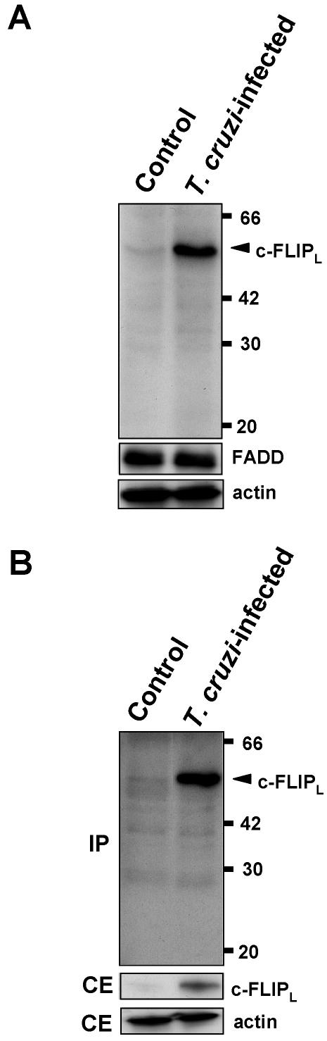
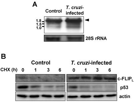
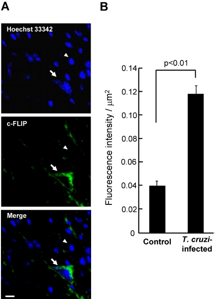
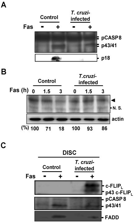
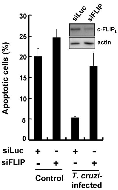
Intracellular persistence of the protozoan parasite, Trypanosoma cruzi, is an aggravating cause of Chagas' disease, involving that the protozoan infection specifically inhibits death receptor-mediated apoptosis of host cells. Here we demonstrate that the parasite dramatically up-regulates cellular FLICE inhibitory protein (c-FLIP), the only known mammalian inhibitor specific for death receptor signaling, in infected cells by an unusual, posttranscriptional stabilization of the short-lived protein. We also show that c-FLIP is accumulated in T. cruzi-infected mouse heart muscle cells in vivo. Stimulation of death receptor Fas in infected cells induces recruitment of c-FLIP to block the procaspase-8 activation at the most upstream caspase cascade. c-FLIP knock-down with a small interfering RNA significantly restores Fas-mediated apoptosis in infected cells. Taken together, our findings indicate that T. cruzi posttranscriptionally up-regulates and exploits host c-FLIP for the inhibition of death-inducing signal, a mechanism that may allow parasites to persist in host cells.


Figures

Figure 1.
Figure 1.
Expression of c-FLIP protein in T. cruzi–infected cells. (A) The cells (2 × 10⁵) were infected with T. cruzi trypomastigotes as described in Materials and Methods. Cell extracts (100 μg protein/lane) were resolved on 12.5% SDS-PAGE, and Western blots were probed with anti-c-FLIP antibody specific for amino acid residues 2–17 and with anti-FADD antibody. Actin was used for loading control. (B) Cell extracts were immunoprecipitated (IP) using anti-c-FLIP mAb (Dave-2). Precipitated samples were analyzed by Western blotting with anti-c-FLIP antibody described in A. Cellular extracts (CE) were also analyzed by Western blotting (10 μg protein/lane). Numbers on the right indicate kilodaltons.
Figure 2.
Figure 2.
Expression of c-FLIP mRNA and effect of cycloheximide treatment. (A) Total RNA (2.5 μg/lane) was resolved on 1% agarose gel, Northern-blotted, and probed for c-FLIP. The band corresponding to c-FLIPL mRNA is indicated by arrowhead. Numbers on the left indicate kilobases. (B) Cells (70–80% confluent) were incubated with 100 μM cycloheximide for the indicated time, and the expression of c-FLIPL and p53 was analyzed by Western blotting. Actin was used for loading control.
Figure 3.
Figure 3.
Immunohistochemical detection of c-FLIP in the cardiomyocytes of mice infected with T. cruzi. (A) Thin sections prepared from T. cruzi–infected mouse hearts were stained with Hoechst 33342 and with anti-c-FLIP mAb (G-11). Details are described in Materials and Methods. Fluorescence images and their merger in the same field in a typical section are shown. The arrow points toward a T. cruzi-dwelling cardiomyocyte. The arrowhead points at a typical uninfected cell. Bar in Merge, 10 μm. (B) The fluorescence intensity of 58 each of T. cruzi–infected and uninfected heart muscle cells was measured for a quantitation of expression level of c-FLIP. Statistical significance was assessed by Student's t test.
Figure 4.
Figure 4.
Processing of procaspase-8 and c-FLIPL in T. cruzi–infected cells. (A) Top: cells were incubated in the presence or absence of anti-Fas antibody for 5 h, and the cell lysates were immunoprecipitated with anti-caspase-8 mAb (12F5). The precipitated protein was analyzed by Western blotting with rabbit anti-caspase-8 polyclonal antibody (GD-13). Bottom: cell extracts (200 μg protein/lane) were fractionated on 12.5% SDS-PAGE and Western-blotted with anti-caspase-8 antibody (GD-13). p18 form processed from p43/41 form of caspase-8 is shown. (B) Cells were stimulated with anti-Fas antibody for 0, 1.5, and 3 h. Each cell extract (55 μg protein/lane) was fractionated on 15% SDS-PAGE and Western-blotted with anti-caspase-8 antibody. Arrowhead indicates the position of pro-caspase-8. Western blots of the panel were scanned using RFLP-scan (Scanalytics, Billerica, MA) and quantified for procaspase-8. The levels of procaspase-8 are shown at the bottom. N.S., nonspecific cross-reactive band. (C) T. cruzi–infected and uninfected cells were stimulated with anti-Fas antibody or were left untreated. After lysis of these cells, DISC or unstimulated Fas was immunoprecipitated by anti-Fas antibody (APO1–3) and analyzed by Western blotting using anti-caspase-8 antibody (GD-13), anti-c-FLIP antibody described in Figure 1A, and anti-FADD antibody. The positions of the proteins and the respective cleavage fragments are indicated.
Figure 5.
Figure 5.
c-FLIP knock-down and recovery of apoptosis in T. cruzi–infected cells. Preparation of siLuc (negative control) and si-FLIP, transfection of these siRNAs into cells, T. cruzi infection, and Fas stimulation were carried out as described in Materials and Methods. siRNA-transfected cells (70–80% confluent), which were subsequently infected with T. cruzi and incubated for 24 h, were stimulated for 5 h with anti-Fas antibody. The percentage of apoptotic cells was determined. In each experiment, more than 350 uninfected or 250 T. cruzi–infected cells were examined. The values are means of three separate experiments and the bars represent standard deviations. The inset shows the expression level of c-FLIPL and actin in T. cruzi–infected cells (15 μg protein/lane). Because of the low expression level of c-FLIPL in control cells, the protein was not detected under the conditions examined.

Similar articles

Cited by

References

    1. Benedict, C. A., Norris, P. S., and Ware, C. F. (2002). To kill or be killed: viral evasion of apoptosis. Nat. Immunol. 3, 1013–1018. - PubMed
    1. Beverley, S. M. (1996). Hijacking the cell: parasites in the driver's seat. Cell 87, 787–789. - PubMed
    1. Brener, Z. (1973). Biology of Trypanosoma cruzi. Annu. Rev. Microbiol. 27, 347–382. - PubMed
    1. Chuenkova, M. V., Furnari, F. B., Cavenee, W. K., and Pereira, M. A. (2001). Trypanosoma cruzi trans-sialidase: a potent and specific survival factor for human Schwann cells by means of phosphatidylinositol 3-kinase/Akt signaling. Proc. Natl. Acad. Sci. USA 98, 9936–9941. - PMC - PubMed
    1. Chuenkova, M. V., and Pereira, M. A. (2000). A trypanosomal protein synergizes with the cytokines ciliary neurotrophic factor and leukemia inhibitory factor to prevent apoptosis of neuronal cells. Mol. Biol. Cell 11, 1487–1498. - PMC - PubMed

Publication types

MeSH terms