. 2005 Oct;73(4):681-7.
doi: 10.1095/biolreprod.104.039164. Epub 2005 May 25.

Conditional induction of ovulation in mice

Affiliations

Conditional induction of ovulation in mice

Pei Su et al. Biol Reprod. 2005 Oct.

Abstract

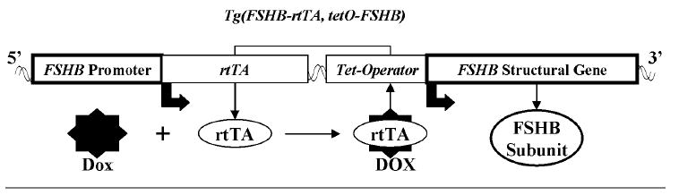
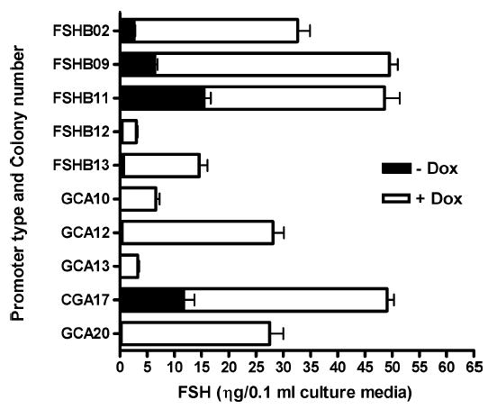
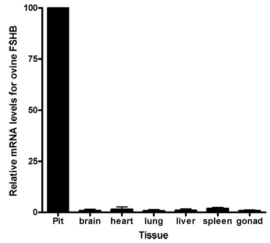
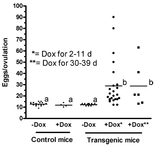
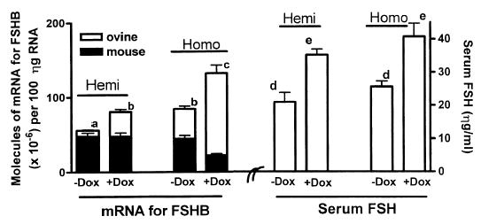
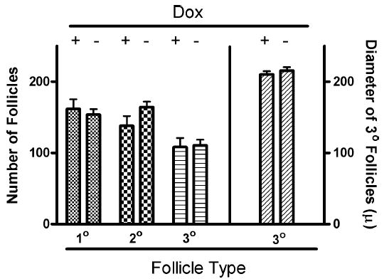
Follicle-stimulating hormone controls the maturation of mammalian ovarian follicles. In excess, it can increase ovulation (egg production). Reported here is a transgenic doxycycline-activated switch, tested in mice, that produced more FSHB subunit (therefore more FSH) and increased ovulation by the simple feeding of doxycycline (Dox). The transgenic switch was expressed selectively in pituitary gonadotropes and was designed to enhance normal expression of FSH when exposed to Dox, but to be regulated by all the hormones that normally control FSH production in vivo. Feeding maximally effective levels of Dox increased overall mRNA for FSHB and serum FSH by over half in males, and Dox treatment more than doubled the normal ovulation rate of female mice for up to 10 reproductive cycles. Lower levels of Dox increased the number of developing embryos by 30%. Ovarian structure and function appeared normal. In summary, gene switch technology and normal FSH regulation were combined to effectively enhance ovulation in mice. Theoretically, the same strategy can be used with any genetic switch to increase ovulation (or any highly conserved physiology) in any mammal.


Figures

FIG. 1
FIG. 1
The ovine Tg(FSHB-rtTA, tetO-FSHB) gene switch. The promoter for ovine FSHB (left box) was used to control expression of the reverse tetracycline-sensitive transactivator (rtTA). This promoter has been shown to express other genes specifically in gonadotropes where they are regulated just like endogenous mouse FSHB subunit protein. Activation of rtTA by doxycycline (Dox), a tetracycline analog, was designed to induce expression of the structural gene for ovine FSHB (right box) by binding to the Tet operator DNA sequence. Because expression of rtTA is designed to mimic expression of endogenous mouse FSHB, Dox activation of rtTA should simply amplify normal regulation of FSHB expression leading to enhanced production of FSHB and FSH in a way that reflects the normal ebb and flow of endogenous FSH.
FIG. 2
FIG. 2
Induction of FSH expression by Dox in LβT2 cells harboring the Tg(CGA-rtTA, tetO-FSHB) or Tg(FSHB-rtTA, tetO-FSHB) gene switches. Five cell lines stably carrying the Tg(CGA-rtTA, tetO-FSHB) gene switch and five cell lines expressing Tg(FSHB-rtTA, tetO-FSHB) were cultured for 48 h with or without 10 μM Dox. The media were assayed by a pan-FSH RIA and the means ± SEMs are shown for triplicate replicates assayed in duplicate for all cell lines. Expression of FSH was significantly increased above control levels for all cell lines (P < 0.001).
FIG. 3
FIG. 3
Pituitary-specific expression of mRNA for ovine FSHB in Tg(FSHB-rtTA, tetO-FSHB)1Wmil mice during Dox treatment. Male mice from Tg(FSHB-rtTA, tetO-FSHB)1Wmil founders were treated with Dox for two weeks, and then tissues were taken and assayed for mRNA of ovine FSHB using real-time RT-PCR. All samples from individual mice were assayed together and the results were normalized to pituitary expression of ‘‘100.’’ To determine differences between several means, analysis of variance was used, followed by the Tukey multiple comparison test for post hoc evaluation of differences between different treatment groups. Assay variation was ≤10%. Individual percentage expressions for brain, heart, lung, liver, spleen, and gonad were 0.9 ± 0.7, 1.5 ± 1.2, 0.7 ± 0.7, 1.0 ± 0.7, 1.9 ± 0.6, and 0.9 ± 0.4, respectively, compared to expression of mRNA for FSHB in the pituitary.
FIG. 4
FIG. 4
Tg(FSHB-rtTA, tetO-FSHB)1Wmil females increased ovulation rates by 240% ± 33% when fed Dox (6g/kg rodent chow) for 2–11 or 30–39 days. Three-month-old hemizygous mice were treated with Dox for 2 or 30 days and then exposed to males. All females showed evidence of copulation (copulation plug) suggesting ovulation within 9 days of exposure to males; >80% showed a plug within 4 days of male exposure. Homozygous mice were also tested at 3 mo of age and showed an ovulation rate of 29 ± 4. Moreover, hemizygous and homozygous mice were tested at age 7 mo with the same increase in ovulation for Dox-fed mice compared to controls. Means of treatment values are designated with a line horizontal to the x-axis. Means that are statistically the same (P < 0.05) have the same letter (a or b).
FIG. 5
FIG. 5
Dox increased total FSH and mRNA for FSHB by 50%–60% in male Tg(FSHB-rtTA, tetO-FSHB)1Wmil mice. No increase was observed in males lacking the fertility switch (not shown). Transgenic B6SJL founder mice were bred into CD-1 mice, giving them considerable CD-1 genetic makeup. Data from the fourth generation are presented in this report. Males were fed rodent chow ± 6g/kg of Dox for 14 days, and then their pituitaries and blood were analyzed. Total RNA was isolated, and mRNAs for ovine FSHB and mouse FSHB were quantified in 100 ηg of RNA using real-time RT-PCR. Standard curves for PCR analyses consisted of known amounts of cDNA for ovine or mouse FSHB in plasmids. Each data point represents the mean ± SEM for either four hemizygous or seven homozygous mice. Statistical analysis used analysis of variance plus the Tukey multiple comparison test. There are significant differences (P < 0.05) between means with different letters.
FIG. 6
FIG. 6
Treatment with Dox for 30–39 days did not alter the number of primary, secondary or tertiary follicles or the size of tertiary follicles (Student t-test; P < 0.05). The data represent the means ± SEM for results from six control and six Dox-treated transgenic mice. The diameter of tertiary follicles is reported in microns.

References

    1. Pursel VG, Bolt DJ, Miller KF, Pinkert CA, Hammer RE, Palmiter RD, Brinster RL. Expression and performance in transgenic pigs. J Reprod Fertil Suppl. 1990;40:235–245. - PubMed
    1. Kumar TR, Palapattu G, Wang P, Woodruff TK, Boime I, Byrne MC, Matzuk MM. Transgenic models to study gonadotropin function: the role of follicle-stimulating hormone in gonadal growth and tumorigenesis. Mol Endocrinol. 1999;13:851–865. - PubMed
    1. Zhu Z, Sheng T, Lee CG, Homer RJ, Elias JA. Tetracycline-controlled transcriptional regulation systems: advances and application in transgenic animal modeling. Semin Cell Dev Bio. 2002;13:121–128. - PubMed
    1. Gossen M, Freundlieb S, Bender G, Muller G, Hillen W, Bujard H. Transcriptional activation by tetracyclines in mammalian cells. Science. 1995;268:1766–1769. - PubMed
    1. Miller CD, Miller WL. Transcriptional repression of the ovine follicle-stimulating hormone-beta gene by 17 beta-estradiol. Endocrinology. 1996;137:3437–3446. - PubMed

Publication types

MeSH terms

Substances