

Large cell neuroendocrine carcinoma of the larynx: a case report and a review of the classification of this neoplasm
- PMID: 15917422
- PMCID: PMC1770703
- DOI: 10.1136/jcp.2004.019927
Large cell neuroendocrine carcinoma of the larynx: a case report and a review of the classification of this neoplasm
Abstract
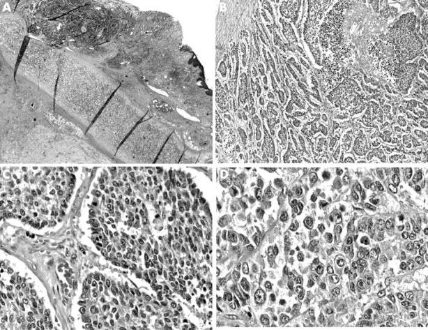
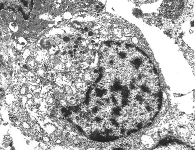
This report describes a case of large cell neuroendocrine carcinoma (LCNEC) of the larynx. A 74 year old man who presented with otalgia underwent direct laryngoscopy with biopsy, which revealed an invasive poorly differentiated carcinoma. Laryngectomy with bilateral neck dissections revealed invasion of the pre-epiglottic space by the tumour, with metastases to bilateral lymph nodes (AJCC T3N2c). The tumour was characterised by large cells with vesicular chromatin and prominent nucleoli. The cells were arranged in organoid and trabecular patterns with a background of extensive necrosis and numerous mitotic figures. Immunohistochemical and ultrastructural analyses confirmed the neuroendocrine nature of the tumour. Metastatic disease was present in the liver, and the patient died within weeks of surgery. LCNEC carcinoma is a rare tumour of the larynx. Recognition at this site is essential so that proper patient management can be initiated.
Figures


Similar articles
-
Large cell neuroendocrine carcinoma of the larynx: definition of an entity.Head Neck Pathol. 2010 Sep;4(3):198-207. doi: 10.1007/s12105-010-0188-0. Epub 2010 Jun 30. Head Neck Pathol. 2010. PMID: 20589486 Free PMC article.
-
Metastatic laryngeal large cell neuroendocrine carcinoma: A rare case of presentation and extreme tumor burden.J Cutan Pathol. 2018 Mar;45(3):229-233. doi: 10.1111/cup.13090. Epub 2017 Dec 28. J Cutan Pathol. 2018. PMID: 29194700
-
Primary combined small cell carcinoma of larynx with lateralized histologic components and corresponding side-specific neck nodal metastasis: report of a unique case and review of literature.Int J Clin Exp Pathol. 2010 Dec 3;4(1):111-7. Int J Clin Exp Pathol. 2010. PMID: 21228933 Free PMC article. Review.
-
Mucosal large cell neuroendocrine carcinoma of the head and neck regions in Japanese patients: a distinct clinicopathological entity.J Clin Pathol. 2012 Aug;65(8):704-9. doi: 10.1136/jclinpath-2012-200801. Epub 2012 Jun 20. J Clin Pathol. 2012. PMID: 22718848 Free PMC article.
-
Typical laryngeal carcinoid tumor with recurrence and lymph node metastasis: a case report and review of the literature.Int J Clin Exp Pathol. 2014 Dec 1;7(12):9028-31. eCollection 2014. Int J Clin Exp Pathol. 2014. PMID: 25674282 Free PMC article. Review.
Cited by
-
Clinico-pathological study of 50 cases of tumours of larynx.Indian J Otolaryngol Head Neck Surg. 2013 Jul;65(Suppl 1):29-35. doi: 10.1007/s12070-011-0420-6. Epub 2011 Dec 13. Indian J Otolaryngol Head Neck Surg. 2013. PMID: 24427612 Free PMC article.
-
Large cell neuroendocrine carcinoma of the larynx: definition of an entity.Head Neck Pathol. 2010 Sep;4(3):198-207. doi: 10.1007/s12105-010-0188-0. Epub 2010 Jun 30. Head Neck Pathol. 2010. PMID: 20589486 Free PMC article.
-
Large cell neuroendocrine carcinoma: topic review and a unique case of metastasis to the mandible.J Maxillofac Oral Surg. 2015 Mar;14(Suppl 1):120-6. doi: 10.1007/s12663-012-0362-x. Epub 2012 Apr 29. J Maxillofac Oral Surg. 2015. PMID: 25861184 Free PMC article.
-
Large cell neuroendocrine carcinoma of the head and neck: a distinct clinicopathologic entity.Eur Arch Otorhinolaryngol. 2014 Aug;271(8):2093-5. doi: 10.1007/s00405-014-3090-7. Epub 2014 May 15. Eur Arch Otorhinolaryngol. 2014. PMID: 24831455 No abstract available.
-
The grey zone between pure (neuro)endocrine and non-(neuro)endocrine tumours: a comment on concepts and classification of mixed exocrine-endocrine neoplasms.Virchows Arch. 2006 Nov;449(5):499-506. doi: 10.1007/s00428-006-0306-2. Epub 2006 Oct 11. Virchows Arch. 2006. PMID: 17033797 Review.
References
-
- Travis W, Brambilla E, Muller-Hermelink H, et al., eds. World Health Organisation classification of tumours. Pathology and genetics of tumours of the lungs, pleura, thymus, and heart. Lyon: IARC Press, 2004.
-
- Evans A, Al-Maghrabi J, Tsihlias J, et al. Primary large cell neuroendocrine carcinoma of the urinary bladder. Arch Pathol Lab Med 2002;126:1229–32. - PubMed
-
- Hailemariam S, Gaspert A, Komminoth P, et al. Primary, pure large-cell neuroendocrine carcinoma of the urinary bladder. Mod Pathol 1998;11:1016–19. - PubMed
-
- Papotti M, Cassoni P, Sapino A, et al. Large cell neuroendocrine carcinoma of the gallbladder. Am J Surg Pathol 2000;24:1424–8. - PubMed
-
- Cavazza A, Gallo M, Valcavi R, et al. Large cell neuroendocrine carcinoma of the ampulla of Vater. Arch Pathol Lab Med 2003;127:221–3. - PubMed
Publication types
MeSH terms
LinkOut - more resources
Full Text Sources