. 2005 Jun-Jul;23(6):781-90.
doi: 10.1634/stemcells.2004-0365.

Directed differentiation of dopaminergic neuronal subtypes from human embryonic stem cells

Affiliations

Directed differentiation of dopaminergic neuronal subtypes from human embryonic stem cells

Yiping Yan et al. Stem Cells. 2005 Jun-Jul.

Abstract

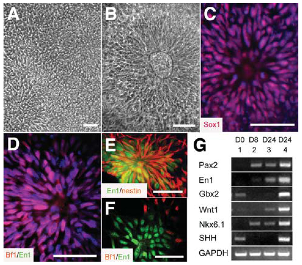
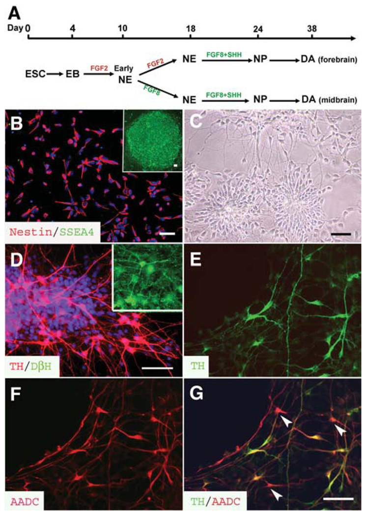
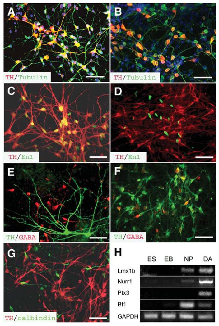
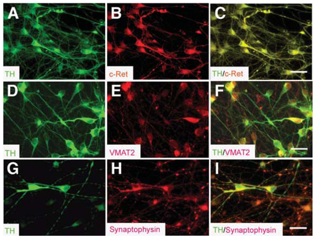
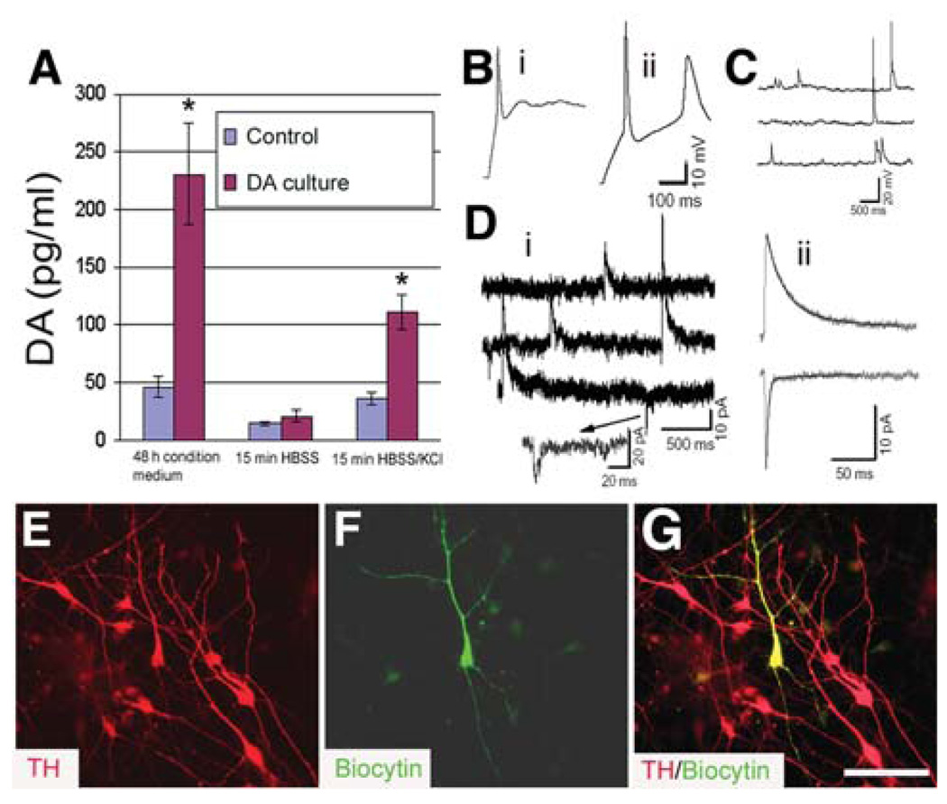
How dopamine (DA) neuronal subtypes are specified remains unknown. In this study we show a robust generation of functional DA neurons from human embryonic stem cells (hESCs) through a specific sequence of application of fibroblast growth factor 8 (FGF8) and sonic hedgehog (SHH). Treatment of hESC-derived Sox1+ neuroepithelial cells with FGF8 and SHH resulted in production of tyrosine hydroxylase (TH)-positive neurons that were mostly bipolar cells, coexpression with gamma-aminobutyric acid, and lack of midbrain marker engrailed 1 (En1) expression. However, FGF8 treatment of precursor cells before Sox1 expression led to the generation of a similar proportion of TH+ neurons characteristic of midbrain projection DA neurons with large cell bodies and complex processes and coexpression of En1. This suggests that one mechanism of generating neuronal subtypes is temporal availability of morphogens to a specific group of precursors. The in vitro-generated DA neurons were electrophysiologically active and released DA in an activity-dependent manner. They may thus provide a renewable source of functional human DA neurons for drug screening and development of sustainable therapeutics for disorders affecting the DA system.


Figures

Figure 1
Figure 1
Specification of midbrain neural progenitors. Columnar cells (A) appeared in the differentiating human embryonic stem cell colony at days 8–10, (B) formed neural tube–like rosettes at day 14, and (C) expressed Sox1. (D): The neuroepithelial cells in FGF2-treated cultures expressed Bf1 (red) but not En1 (green). (E): En1 (green) expression was observed in the nestin+ (red) neural progenitors after 6 days of treatment with FGF8 (100 ng/ml) at day 10, expansion in FGF8 for 2 days, and subsequent treatment with FGF8 and SHH (200 ng/ml) for another 6 days on laminin substrate. (F): These En1+ cells (green) were negative for Bf1 (red). The cell nuclei were stained with Hoechst (bluein C, D). Bar = 50 µm. (G): Reverse transcription–polymerase chain reaction showed a higher expression level of Pax2, En1, Wnt1, and Gbx2, as well as Nkx6.1 and SHH mRNAs, when the embryonic stem cell (lane 1)–derived early neuroepithelial cells (lane 2) were treated with FGF8 followed by FGF8 and SHH (lane 4) compared with the cells that were treated with FGF2 followed by FGF8 and SHH for the same period (lane 3). Abbreviations: FGF, fibroblast growth factor; SHH, sonic hedgehog.
Figure 2
Figure 2
Differentiation of DA neurons. (A): Schematic procedures of DA neuron differentiation. hESCs were differentiated to early neuroepithelial cells in the form of early rosettes at 8–10 days, followed by formation of neural tube–like rosettes for another week in the presence of either FGF2 (late FGF8 treatment) or FGF8 (early FGF8 treatment). They were then differentiated to NPs in the presence of FGF8 and SHH for 6 days before proceeding to DA neurons in the next 2–3 weeks after withdrawal of morphogens. (B): The neural progenitors derived form hESCs were positive for nestin (red) and negative for SSEA4 (green), which was expressed in the undifferentiated hESCs (inset). (C): The dissociated NPs reformed rosettes and began to extend neurorites 5 days after plating. (D): Approximately one third of the differentiated cells were TH+ (red) but DβH (green) in the early FGF8 treatment group at 3 weeks of differentiation. The inset indicates that DβH positively stained cells in the section of adult rat brainstem. (E–G): All TH+ cells (E, green) were positively stained with AADC (F and G, red), but some AADC+ cells were negative for TH (G, arrowheads). The cell nuclei were stained with Hoechst (B and D, blue). Bar = 50 µm. Abbreviations: A ADC, amino acid decarboxylase; DA, dopamine; EB, embryoid body; ESC, embryonic stem cell; FGF, fibroblast growth factor; hESC, human embryonic stem cell; NE, neuroepithelia; NP, neural progenitor; SHH, sonic hedgehog; SSEA, stage-specific embryonic antigen; TH, tyrosine hydroxylase.
Figure 3
Figure 3
Characterization of human ES cell–derived DA neurons. (A, B): All TH+ cells (red) in the (A) early and (B) late FGF8-treated cultures were positively stained for βIII-tubulin (green). The TH+ cells exhibited (A) multipolar morphology in the early FGF8 treatment culture and (B) bipolar morphology in the late FGF8 treatment culture. (C, D): Coexpression of TH+ (red) and En1 (green) was observed in (C) the early FGF8 treatment cultures but not in (D) the late FGF8 treatment culture. (E, F): Few TH+ neurons (green) coexpressed GABA (red) in the (E) early FGF8 treatment cultures compared with the (F) late FGF8 treatment group. (G): The TH+ cells (red) in the early FGF8 treatment culture were negative for calbindin (green). The cell nuclei were stained with Hoechst (A and B, blue). Bar = 50 µm. (H): Reverse transcription–polymerase chain reaction analyses indicated the expression of other dopaminergic-related genes when the ES cells were differentiated into DA neurons through EB and NP stages. Abbreviations: DA, dopamine; EB, embryoid body; ES, embryonic stem; FGF, fibroblast growth factor; GABA, γ-aminobutyric acid; NP, neural progenitor; TH, tyrosine hydroxylase.
Figure 4
Figure 4
Expression of receptors and transporters in the human embryonic stem cell–derived midbrain dopamine neurons. (A–C): All TH+ cells (A, green) expressed c-Ret (B and C, red). (D–F): TH+ cells (D, green) coexpressed VMAT2 (E and F; red). (G–I): TH+ neurons (G, green) expressed synaptophysin (H and I, red). Bar = 25 µm. Abbreviations: TH, tyrosine hydroxylase; VMAT, vesicular monoamine transporter.
Figure 5
Figure 5
Functional characteristics of the in vitro–generated midbrain DA neurons. (A): Spontaneous and depolarization (56 mM KCl in HBSS)–induced DA release in the non-DA cultures and the early fibroblast growth factor 8–treated cultures at 3 weeks of differentiation. Data were presented as mean ± SD from three experiments. *p < .05 versus control by the unpaired Student’s t-test. (B): Action potentials evoked by depolarizing current steps (0.2 nA) in two neurons differentiated for 30 days. Passive membrane properties: (i) Vrest −49 mV, Cm 15.5 pF, Rm 5.0 GΩ; (ii) Vrest −72 mV, Cm 45 pF, Rm 885 MΩ. (C): Spontaneous postsynaptic potentials in a neuron differentiated for 36 days. (D): Spontaneous postsynaptic currents in a neuron differentiated for 30 days in culture. The neuron was voltage clamped at −40 mV using a K gluconate–based pipette solution. The outward currents reflect inhibitory events, and the inward currents reflect excitatory events in this low-chloride recording solution. (ii): Averaged events from the cell illustrated in panel (i). The weighted decay time constants are 61.4 ms and 9.9 ms for inhibitory (n = 17 events) and excitatory (n = 14 events) currents, respectively. (E–G): Immunostaining showed that the recorded neuron (F, green) was positive for TH (E and G, red). Bar = 50 µm. Abbreviations: DA, dopamine; HBSS, Hanks’ balanced salt solution; TH, tyrosine hydroxylase.

References

    1. Bjorklund A, Lindvall O. Dopamine-containing systems in the CNS. In: Bjorklund A, Hokfelt T, editors. Handbook of Chemical Neuroanatomy. Vol. 2. Amsterdam: Elsevier Science publishers; 1984. pp. 55–111. Classical Transmitters in the CNS.
    1. Gall CM, Hendry SH, Seroogy KB, et al. Evidence for coexistence of GABA and dopamine in neurons of the rat olfactory bulb. J Comp Neurol. 1987;266:307–318. - PubMed
    1. Kosaka T, Kosaka K, Hataguchi Y, et al. Catecholaminergic neurons containing GABA-like and/or glutamic acid decarboxylase-like immunore-activities in various brain regions of the rat. Exp Brain Res. 1987;66:191–210. - PubMed
    1. Zetterstrom RH, Solomin L, Jansson L, et al. Dopamine neuron agenesis in Nurr1-deficient mice. Science. 1997;276:248–250. - PubMed
    1. Smidt MP, van Schaick HS, Lanctot C, et al. A homeodomain gene Ptx3 has highly restricted brain expression in mesencephalic dopaminergic neurons. Proc Natl Acad Sci U S A. 1997;94:13305–13310. - PMC - PubMed

Publication types

MeSH terms