Comparative Study
. 2005 Jun;49(6):2438-44.
doi: 10.1128/AAC.49.6.2438-2444.2005.

Comparable efficacies of the antimicrobial peptide human lactoferrin 1-11 and gentamicin in a chronic methicillin-resistant Staphylococcus aureus osteomyelitis model

Affiliations
Comparative Study

Comparable efficacies of the antimicrobial peptide human lactoferrin 1-11 and gentamicin in a chronic methicillin-resistant Staphylococcus aureus osteomyelitis model

Christopher Faber et al. Antimicrob Agents Chemother. 2005 Jun.

Abstract

The therapeutic efficacy of an antimicrobial peptide, human lactoferrin 1-11 (hLF1-11), was investigated in a model of chronic methicillin-resistant Staphylococcus aureus (MRSA) (gentamicin susceptible) osteomyelitis in rabbits. We incorporated 50 mg hLF1-11/g or 50 mg gentamicin/g cement powder into a calcium phosphate bone cement (Ca-P) and injected it into the debrided tibial cavity, creating a local drug delivery system. The efficacy of hLF1-11 and gentamicin was compared to that of a sham-treated control (plain bone cement) (n=6) and no treatment (infected only) (n=5). The results were evaluated by microbiology, radiology, and histology. MRSA was recovered from all tibias in both control groups (n=11). On the other hand, hLF1-11 and gentamicin significantly reduced the bacterial load. Furthermore, no growth of bacteria was detected in five out of eight and six out of eight specimens of the hLF1-11- and gentamicin-treated groups, respectively. These results were confirmed by a significant reduction of the histological disease severity score by hLF1-11 and gentamicin compared to both control groups. The hLF1-11-treated group also had a significantly lower radiological score compared to the gentamicin-treated group. This study demonstrates the efficacy of hLF1-11 incorporated into Ca-P bone cement as a possible therapeutic strategy for the treatment of osteomyelitis, showing efficacy comparable to that of gentamicin. Therefore, the results of this study warrant further preclinical investigations into the possibilities of using hLF1-11 for the treatment of osteomyelitis.


Figures

FIG. 1.
FIG. 1.
Preoperative lateral radiograph (A) of the right tibia from a sham-treated rabbit. Periosteal reaction is present (arrowheads), and the drill hole can still be seen as a slight shadow (arrow). The postoperative lateral radiograph (B) of the same rabbit shows the cement in situ (arrows).
FIG. 2.
FIG. 2.
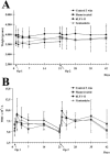
Changes in body weight and white blood cell (WBC) count are plotted over time in A and B, respectively. The first and second surgical procedure are indicated as OP1 and OP2, respectively. There were no significant differences between groups. Values are the means ± standard deviation.
FIG. 3.
FIG. 3.
Quantitative microbiological analyses of the cultured bone homogenate. The individual data points (circles) and the medians (black bars) are indicated. The dotted line represents the approximate detection limit (≈2.5 × 10³ CFU/g bone) of the assay; animals with values below this limit had negative cultures with the quantitative method (no growth). Both the 3-week control and the sham-treated animals had a significantly higher bacterial load than the hLF1-11- (P = 0.01) or gentamicin-treated animals (P = 0.004). There was no significant difference in bacterial load between the 3-week controls and the sham-treated animals (P = 0.6), nor was there a difference between the gentamicin- and hLF1-11-treated animals (P = 0.6).
FIG. 4.
FIG. 4.
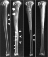
Representative radiographs of excised tibias. In the 3-week control animal (A), periosteal reaction is visible (white arrowheads). More severe signs of chronic osteomyelitis are seen in the 6-week sham-treated animal (B). The periosteal reaction (arrowheads) is exacerbated and a sequestrum is present (arrows). Also, there is a moth-eaten appearance with bony destruction (stars). More proximal, there is evidence of bone cement resorption (block arrows). Slight periosteal elevation (arrowheads) was seen in the gentamicin-treated tibia (C) distal to the bone cement, whereas this hLF1-11-treated animal (D) did not show signs of osteomyelitis.
FIG. 5.
FIG. 5.
Radiographic osteomyelitis scoring per Norden et al. was used to analyze disease severity at day 21 (white bars) and day 42 (black bars). The maximum score of 7 reflects severe chronic osteomyelitis in the entire tibia with periosteal new bone, sequestra, and foci of necrosis. The disease severity score increased from day 21 to day 42 in both the sham-treated group (P = 0.04) and the gentamicin group (P = 0.011). Furthermore, at day 42, the hLF1-11-treated animals had a significantly better score than both the sham-treated (P = 0.043) and the gentamicin-treated animals (P = 0.035). Given are the mean values and standard deviation.
FIG. 6.
FIG. 6.
Representative photomicrographs (hematoxylin and eosin stained) of transverse sections of the tibial cortex. Panel A depicts a 3-week control animal; note the marked periosteal reaction (stars) surrounding the largely necrotic cortex. Also, abscesses (white arrows) are present both intramedullarly and subperiosteally. The intramedullary canal is filled with fibrotic tissue (arrowheads) and some new bone formation also can be seen (black arrow). Panel B depicts a sham-treated specimen. Note the bone marrow versus bone cement interface (white block arrows). Similar pathology can be seen with periosteal bone formation (stars), abscesses (white arrows), intramedullary fibrosis (arrowheads), and intramedullary new bone formation (black arrows). Panel C shows an hLF1-11-treated specimen. Note the absence of periosteal inflammation (white arrows); no other pathology can be seen. A gentamicin-treated specimen without periosteal inflammation (white arrows) is depicted in panel D. Magnification: A and B, 25×; C and D, 50×.
FIG. 7.
FIG. 7.
Histopathological data as scored using the score of Smeltzer et al. (mean + standard deviation). The maximum score is 16, indicating severe osteomyelitis with intramedullary abscess and fibrosis, severe periosteal reaction with subperiosteal abscess, and multiple foci of necrotic bone. Both the hLF1-11- and gentamicin-treated animals have a significantly lower score than the 3-week control and the sham groups, P = 0.04 and P = 0.01, respectively.

References

    1. Castro, C., E. Sanchez, A. Delgado, I. Soriano, P. Nunez, M. Baro, A. Perera, and C. Evora. 2003. Ciprofloxacin implants for bone infection. In vitro-in vivo characterization. J. Control. Release 93:341-354. - PubMed
    1. Craig, W. A. 1995. Once-daily versus multiple-daily dosing of aminoglycosides. J. Chemother. 7(Suppl. 2):47-52. - PubMed
    1. Giacometti, A., O. Cirioni, R. Ghiselli, F. Mocchegiani, M. S. Del Prete, C. Viticchi, W. Kamysz, E. LEmpicka, V. Saba, and G. Scalise. 2002. Potential therapeutic role of cationic peptides in three experimental models of septic shock. Antimicrob. Agents Chemother. 46:2132-2136. - PMC - PubMed
    1. Hancock, R. E. 1997. Peptide antibiotics. Lancet 349:418-422. - PubMed
    1. Hill, J., L. Klenerman, S. Trustey, and R. Blowers. 1977. Diffusion of antibiotics from acrylic bone-cement in vitro. J. Bone Joint Surg. Br. 59:197-199. - PubMed

Publication types

MeSH terms