. 2005 Jun;49(6):2445-53.
doi: 10.1128/AAC.49.6.2445-2453.2005.

Antifungal protein PAF severely affects the integrity of the plasma membrane of Aspergillus nidulans and induces an apoptosis-like phenotype

Affiliations
Free PMC article

Antifungal protein PAF severely affects the integrity of the plasma membrane of Aspergillus nidulans and induces an apoptosis-like phenotype

Eva Leiter et al. Antimicrob Agents Chemother. 2005 Jun.
Free PMC article

Abstract

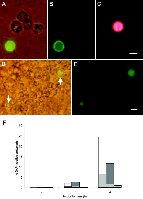
The small, basic, and cysteine-rich antifungal protein PAF is abundantly secreted into the supernatant by the beta-lactam producer Penicillium chrysogenum. PAF inhibits the growth of various important plant and zoopathogenic filamentous fungi. Previous studies revealed the active internalization of the antifungal protein and the induction of multifactorial detrimental effects, which finally resulted in morphological changes and growth inhibition in target fungi. In the present study, we offer detailed insights into the mechanism of action of PAF and give evidence for the induction of a programmed cell death-like phenotype. We proved the hyperpolarization of the plasma membrane in PAF-treated Aspergillus nidulans hyphae by using the aminonaphtylethenylpyridinium dye di-8-ANEPPS. The exposure of phosphatidylserine on the surface of A. nidulans protoplasts by Annexin V staining and the detection of DNA strand breaks by TUNEL (terminal deoxynucleotidyltransferase-mediated dUTP-biotin nick end labeling) gave evidence for a PAF-induced apoptotic-like mechanism in A. nidulans. The localization of reactive oxygen species (ROS) by dichlorodihydrofluorescein diacetate and the abnormal cellular ultrastructure analyzed by transmission electron microscopy suggested that ROS-elicited membrane damage and the disintegration of mitochondria played a major role in the cytotoxicity of PAF. Finally, the reduced PAF sensitivity of A. nidulans strain FGSC1053, which carries a dominant-interfering mutation in fadA, supported our assumption that G-protein signaling was involved in PAF-mediated toxicity.


Figures

FIG. 1.
FIG. 1.
Effect of mutations affecting G-protein signaling in A. nidulans. (A) Conidia of the strains FGSC33, RJH046 (ΔflbA), FGSC116, and FGSC1035 (fadAG203R) were treated with 10 μg of PAF/ml for 48 h at room temperature. (B) Fungal hyphae were pretreated with 100 μM concentrations of the nucleotide analogs GTPγS (▪), GDPβS (□), and ATPγS (░⃞) before 10 μg of PAF/ml was added, followed by incubation for 24 h at room temperature. Values represent the growth percentage of the tested strains compared to the untreated control (i.e., 100%). Samples were prepared in triplicates.
FIG. 2.
FIG. 2.
Potentiometric fluorescence staining of A. nidulans wt hyphae with di-8-ANEPPS. (A to C) The plasma membrane of A. nidulans was loaded with di-8-ANEPPS for 20 min prior to PAF addition. (D to F) Hyperpolarization of the cellular membranes reached its maximum after 80 min of incubation with 10 μg of PAF/ml at room temperature. The fluorescence was recorded at 560 nm (A and D) and at 620 nm (B and E) and micrographs were merged (C and F). Arrows indicate hyphal tips. Scale bar, 5 μm.
FIG. 3.
FIG. 3.
Fluorescence intensity difference of di-8-ANEPPS labeled and PAF-treated A. nidulans wt hyphae as a function of incubation time. (A) Hyperpolarization of the cellular membranes, as assessed by calculating the ratios of images representing ROIs, reached its maximum after 80 min of incubation with 10 μg of PAF/ml at room temperature. Ratio images were obtained at 1, 40, and 80 min after the addition of PAF. (B) Differences in intensity ratios of F620/F560 determined near the first septum and at the hyphal tip over the 80 min of incubation with 10 μg of PAF/ml at room temperature. The results are expressed as mean ± the standard deviation (n = 3). Scale bar, 25 μm.
FIG. 4.
FIG. 4.
Detection of intracellular ROS by H2DCFDA in A. nidulans wt. (A and B) Hyphae were treated with 50 μg of PAF/ml for 90 min at room temperature (A) or with 5 μg of nystatin/mg (hyphal wet weight) as a control for ROS generation (B) before labeling with H2DCFDA. (C) Untreated control labeled with H2DCFDA. (D) Phase contrast of image in panel C. Scale bar, 10 μm.
FIG. 5.
FIG. 5.
Detection of apoptotic markers in A. nidulans protoplasts after treatment with 50 μg of PAF/ml. Annexin V (A and B)- and PI (C)-stained protoplasts after PAF exposure for 3 h and TUNEL-stained nuclei in protoplasts after incubation for 1 h (D and E) were visualized by microscopy by using phase contrast and fluorescence (A and D) and fluorescence alone (B, C, and E), respectively. Two TUNEL-positive cells are indicated by arrows in panel D. Scale bars, 5 μm. (F) Results are shown as the percentage of DAPI-positive protoplasts that stain positive with Annexin V (white), TUNEL (dark gray), or PI (black) at time zero and after 1 and 3 h of incubation with PAF. Light gray portions indicate the percentage of positively stained protoplasts without PAF treatment.
FIG. 6.
FIG. 6.
TEM images of the cellular ultrastucture of A. nidulans. (A to C) Untreated control; (D to F) specimens were treated with 50 μg of PAF/ml for 3 h at room temperature. CW, cell wall; ER, endoplasmic reticulum; G, glycogen; M, mitochondrion; MVS, microvesicular structures; N, nucleus; n, nucleolus; P, peroxisome; PM, plasma membrane; R, ribosomes; V, vacuole. Scale bars: 0.25 μm (A, B, C, E, and F) and 0.6 μm (D).

Similar articles

Cited by

References

    1. Adams, T. H., J. Wieser, and J.-H. Yu. 1998. Asexual sporulation in Aspergillus nidulans. Microbiol. Mol. Biol. Rev. 62:35-54. - PMC - PubMed
    1. Arends, M. J., R. G. Morris, and A. H. Wyllie. 1990. Apoptosis: the role of the endonuclease. Am. J. Pathol. 136:593-608. - PMC - PubMed
    1. Arndt-Jovin, D. J., and T. M. Jovin. 1989. Fluorescence labeling and microscopy of DNA. Methods Cell Biol. 30:417-448. - PubMed
    1. Barratt, R. W., G. B. Johnson, and W. N. Ogata. 1965. Wild-type and mutant stocks of Aspergillus nidulans. Genetics 52:233-246. - PMC - PubMed
    1. Beach, J. M., E. D. McGahren, J. Xia, and B. R. Duling. 1996. Ratiometric measurement of endothelial depolarization in arterioles with a potential-sensitive dye. Am. J. Physiol. 270:2216-2227. - PubMed

Publication types

MeSH terms

LinkOut - more resources