. 2005 Aug 1;566(Pt 3):865-84.
doi: 10.1113/jphysiol.2005.089664. Epub 2005 May 26.

A functional glutamatergic neurone network in the medial septum and diagonal band area

Affiliations

A functional glutamatergic neurone network in the medial septum and diagonal band area

F Manseau et al. J Physiol. .

Abstract

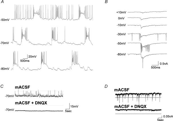
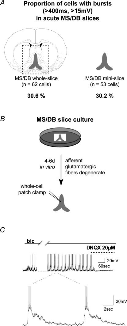
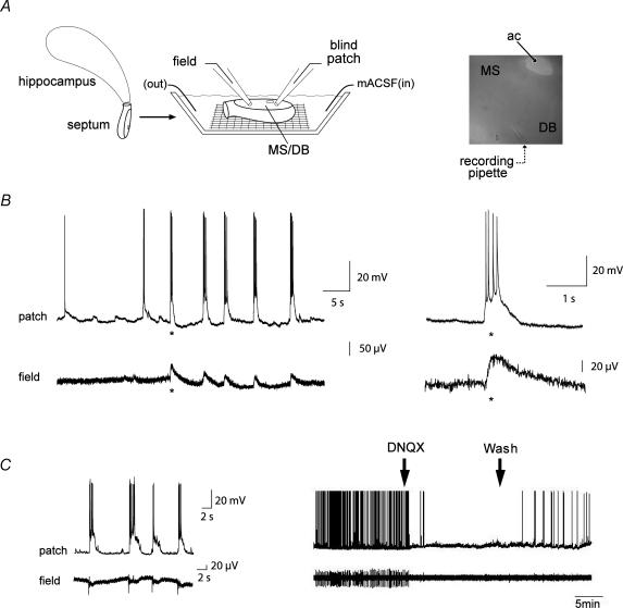
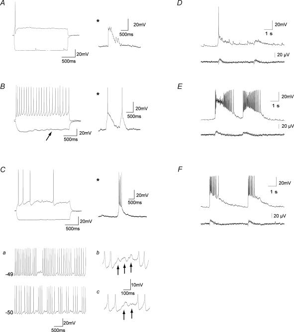
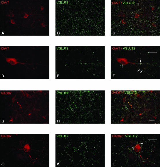
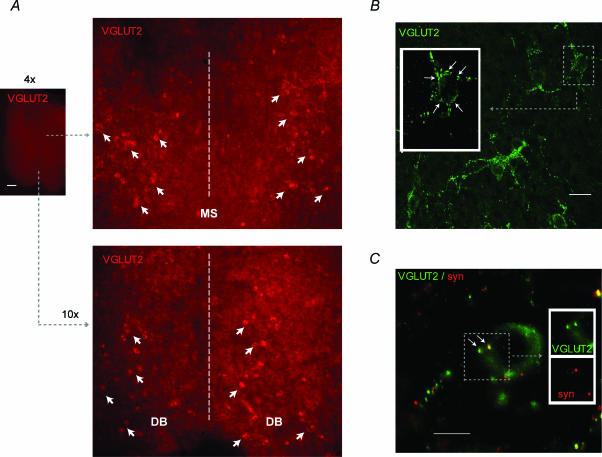
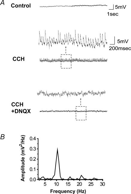
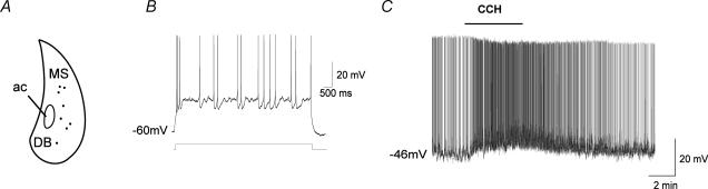
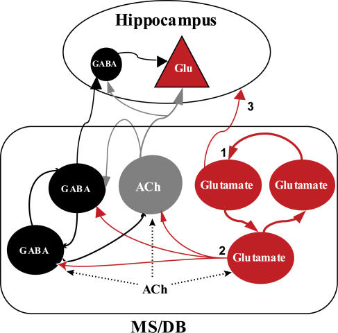
The medial septum and diagonal band complex (MS/DB) is important for learning and memory and is known to contain cholinergic and GABAergic neurones. Glutamatergic neurones have also been recently described in this area but their function remains unknown. Here we show that local glutamatergic neurones can be activated using 4-aminopyridine (4-AP) and the GABA(A) receptor antagonist bicuculline in regular MS/DB slices, or mini-MS/DB slices. The spontaneous glutamatergic responses were mediated by AMPA receptors and, to a lesser extend, NMDA receptors, and were characterized by large, sometimes repetitive activity that elicited bursts of action potentials postsynaptically. Similar repetitive AMPA receptor-mediated bursts were generated by glutamatergic neurone activation within the MS/DB in disinhibited organotypic MS/DB slices, suggesting that the glutamatergic responses did not originate from extrinsic glutamatergic synapses. It is interesting that glutamatergic neurones were part of a synchronously active network as large repetitive AMPA receptor-mediated bursts were generated concomitantly with extracellular field potentials in intact half-septum preparations in vitro. Glutamatergic neurones appeared important to MS/DB activation as strong glutamatergic responses were present in electrophysiologically identified putative cholinergic, GABAergic and glutamatergic neurones. In agreement with this, we found immunohistochemical evidence that vesicular glutamate-2 (VGLUT2)-positive puncta were in proximity to choline acetyltransferase (ChAT)-, glutamic acid decarboxylase 67 (GAD67)- and VGLUT2-positive neurones. Finally, MS/DB glutamatergic neurones could be activated under more physiological conditions as a cholinergic agonist was found to elicit rhythmic AMPA receptor-mediated EPSPs at a theta relevant frequency of 6-10 Hz. We propose that glutamatergic neurones within the MS/DB can excite cholinergic and GABAergic neurones, and that they are part of a connected excitatory network, which upon appropriate activation, may contribute to rhythm generation.


Figures

Figure 1
Figure 1. Large spontaneous glutamatergic bursts are observed in neurones from acute MS/DB slices perfused with mACSF (4-AP, bicuculline and Mg2+free)
A, prolonged perfusion (more than 30 min) with mACSF induced spontaneous and sustained bursting discharges in a MS/DB neurone. Example from a cell that was recorded for more than 1 h, 72 min after the start of mACSF perfusion, demonstrating large and long-lasting bursts (mean duration, 1071 ± 64.4 ms; mean amplitude, 35 ± 2.3 mV; n = 19 bursts) at regular intervals. Current-clamping the cell at different membrane potentials did not affect the regularity of bursts, indicating that this activity was not due to intrinsic membrane properties but rather to synaptic input. Note that spontaneous bursts can trigger action potentials from a holding potential of −80 mV. B, large spontaneous inward currents recorded under voltage clamp from the neurone shown in A have an equilibrium potential typical of glutamatergic transmission (> 0 mV). C, for a different neurone, upper and lower traces illustrate current-clamp recordings (duration, 1 min) of mACSF-induced spontaneous activity before and after the addition of DNQX (in this example, bursts did not trigger action potentials from the holding potential of −80 mV). In current-clamp recorded MS/DB neurones, the mACSF-induced bursts were abolished by the AMPA/kainate receptor antagonist DNQX (20 μm, *P < 0.05, paired t test, n = 5). D, in voltage-clamp recordings of the same cells (held at −80 mV), mACSF-induced spontaneous inward currents exceeding a preset threshold (horizontal dashed line shows three times the baseline noise) were also completely abolished by DNQX (**P < 0.01, n = 5).
Figure 2
Figure 2. AMPA receptor-mediated bursts are prominent in MS/DB acute and organotypic mini-slices containing only the MS/DB
A, narrowing the slice preparation to the MS/DB region by surgically removing the neighbouring lateral septum did not reduce the proportion of cells showing large spontaneous EPSPs (measured from a holding of –80 mV). The contours of the complete (regular) slice are outlined (dashed line) and include the lateral septum and portions of the striatum. B, organotypic slices containing the MS/DB region alone were cultured for 4– 6 days in vitro to allow afferent (extra-septal) glutamatergic terminals to degenerate. C, disinhibition of the organotypic slice with bicuculline rapidly (in less than 5 min) induced large spontaneous and repetitive bursting discharges that were abolished by DNQX (20 μm, n = 6). The bottom panel shows an expanded view of the spontaneous bursts at a faster sweep speed.
Figure 3
Figure 3. Synchronized intracellular glutamate-mediated bursts and extracellular field potentials in a novel in vitro half-septum preparation
A, schematic diagram of the recording set up. Field and patch electrodes were gently lowered onto the exposed MS/DB surface of the half-septa, and whole-cell recordings were established using the blind patch technique. The image on the right illustrates the positioning of a recording electrode in the centre of the MS with the anterior commissure (ac) serving as a landmark. The picture was taken at 10 × magnification. B, spontaneous repetitive bursts were recorded in MS/DB neurones (top traces) and were correlated with large field potentials (bottom traces) in the mACSF-infused half-septum. Expanded traces (right, *) show that the extracellular bursts occur synchronously with those in the cell. C, in another experiment, the spontaneous intra- and extracellular bursting activity observed in mACSF (shown here at rapid sweep speed on the left and slow sweep speed on the right) were reversibly blocked by the AMPA receptor antagonist DNQX (20 μm). These results indicate that MS/DB glutamatergic neurones are organized into a network. For this experiment, the tip of the extracellular electrode was placed 200 μm form the recorded neurone.
Figure 4
Figure 4. Electrophysiologically identified MS/DB neurones receive glutamatergic input in mACSF-perfused MS/DB slices and display synchronized glutamatergic network activity in the intact half-septum preparation
AC, neurones challenged with depolarizing and hyperpolarizing currents (left traces) to identify cell types and examples of burst observed in these are shown (*). Large excitatory bursts (*) were observed in slow-firing (putative cholinergic) neurones showing no Ih current, strong spike frequency accommodation and a large AHP (A), fast-firing (putative GABAergic) neurones showing prominent Ih current (arrow), nominal accommodation and small AHP (B) and cluster-firing (putative glutamatergic) neurones of the MS/DB (C). C, examples of cluster-firing (a) and subthreshold oscillations (b and c) showing increased frequency at depolarized membrane potentials are shown on the right for each trace depicted in a. DF, representative examples of repetitive bursts (upper traces) and synchronized field potentials (lower traces) in slow- (D), fast- (E) and cluster-firing (F) neurones.
Figure 5
Figure 5. Immunohistochemical evidence for locally originating VGLUT2-positive terminals on MS/DB cholinergic and GABAergic neurones in organotypic slices
AF, representative examples showing (AC) numerous VGLUT2-positive puncta (green) in a region containing many ChAT-positive neurones (red) and (DF) VGLUT2-positive terminals (green; arrows) in contact with the soma and dendrites of a ChAT-positive neurone (red). Single-channel confocal immunofluorescence for ChAT (A and D), VGLUT2 (B and E) and merged images (C and F). GL, representative examples showing (GI) numerous VGLUT2-positive puncta (green) in a region containing many GAD67-positive neurones (red) and (JL) VGLUT2-positive terminals (green; arrows) in contact with the soma and dendrites of a GAD67-positive neurone (red). Single-channel confocal immunofluorescence for GAD67 (G and J), VGLUT2 (H and K) and merged images (I and L). Note that although the putative glutamatergic synapses seem to be relatively scattered in the tissue, a clear correspondence of VGLUT2 distribution with the dendritic pattern of the labelled neurones is seen in the preceding examples. Scale bars, 20 μm.
Figure 6
Figure 6. Fluorescence microscopy (A) and confocal (B and C) photomicrographs of VGLUT2-positive neurones and terminals in colchicine-treated organotypic MS/DB slices
A, VGLUT2-immunostaining following a short (1-h) exposure to colchicine reveals the presence of numerous Alexa Fluor 568-positive (glutamatergic) cell bodies (red; arrows) near the lateral borders of the MS and throughout the diagonal bands on the ventral side of the slice (dotted line, midline). A similar distribution was seen in eight other slices. B, Alexa Fluor 488-labelled VGLUT2 puncta and neurones (green). The VGLUT2-positive neurones were often organized in elongated clusters as illustrated here and many seemed surrounded with VGLUT2-positive terminals. Inset, higher magnification from a few optical sections of the area outlined in B illustrating putative VGLUT2-positive teminals (arrows) lining the surface of a VGLUT2-positive soma. C, Alexa Fluor 488-labelled VGLUT2 neurone (green) and colocalization of VGLUT2-positive puncta (green) with Cy3-labelled synaptophysin puncta (red). Inset, higher magnification of area outlined in C illustrating VGLUT2/synaptophysin-postive terminals (arrows) contacting the VGLUT2-positive neurone. Number of stacked optical sections for B and C are 16 and 6, respectively. Scale bars: 500 μm in A; 20 μm in B; and 10 μm in C.
Figure 7
Figure 7. Selective activation of muscarinic receptors elicits large and rhythmic EPSPs in MS/DB neurones in the in vitro half-septum preparation
A, in control (ACSF) solution, no apparent synaptic activity is seen in a whole-cell recorded neurone of the half-septum preparation (top trace; membrane potential, −55 mV). However, bath application of the cholinergic agonist carbachol (CCH, 30 μm) induces continuous and rhythmic EPSPs (middle trace), which are abolished by the addition of the glutamatergic AMPA/kainate receptor antagonist DNQX (20 μm, bottom trace). B, power spectrum of carbachol-induced synaptic activity obtained over a 2-s recording from the neurone shown in A using a fast Fourier transform algorithm (pCLAMP 9.0). Note the distinct peak at 10 Hz.
Figure 8
Figure 8. Distribution of the recorded cluster-firing (putative glutamatergic) neurones in the intact half-septum preparation and depolarizing effect of carbachol
A, schematic diagram showing the approximate location of cluster-firing cells recorded from the MS/DB area in 12 preparations. B, current-clamp recording showing the response of a cluster-firing MS/DB neurone to injection of a depolarizing current pulse (8-s square pulse) applied from a membrane potential of −60 mV. Depolarization elicited characteristic cluster firing and subthreshold oscillations between clusters. C, in the same cell, bath application of carbachol (30 μm) caused a 7 mV depolarization from resting potential and dramatically increased the firing frequency (from 50 to a 100 action potentials min−1) in a reversible manner.
Figure 9
Figure 9. Simplified model of interactions between MS/DB neurones and hippocampus
In this model, glutamatergic neurones are part of a network (1) and can activate (2) GABAergic and cholinergic neurones mainly through AMPA-type receptors. Some glutamatergic neurones (Sotty et al. 2003) may be septohippocampal-projecting neurones (3). Acetylcholine activates GABAergic, cholinergic and glutamatergic neurones of the MS/DB through muscarinic receptors.

Similar articles

Cited by

References

    1. Alonso A, Gaztelu JM, Buno W, JR, Garcia-Austt E. Cross-correlation analysis of septohippocampal neurons during theta-rhythm. Brain Res. 1987;413:135–146. - PubMed
    1. Alreja M, Wu M, Liu W, Atkins JB, Leranth C, Shanabrough M. Muscarinic tone sustains impulse flow in the septohippocampal GABA but not cholinergic pathway: implications for learning and memory. J Neurosci. 2000;20:8103–8110. - PMC - PubMed
    1. Amaral DG, Kurz J. An analysis of the origins of the cholinergic and noncholinergic septal projections to the hippocampal formation of the rat. J Comp Neurol. 1985;240:37–59. - PubMed
    1. Armstrong JN, MacVicar BA. Theta-frequency facilitation of AMPA receptor-mediated synaptic currents in the principal cells of the medial septum. J Neurophysiol. 2001;85:1709–1718. - PubMed
    1. Avoli M, D'Antuono M, Louvel J, Kohling R, Biagini G, Pumain R, D'Arcangelo G, Tancredi V. Network and pharmacological mechanisms leading to epileptiform synchronization in the limbic system in vitro. Prog Neurobiol. 2002;68:167–207. - PubMed

Publication types

LinkOut - more resources