. 2005 Jun 7;102(23):8180-5.
doi: 10.1073/pnas.0408867102. Epub 2005 May 26.

Electrostatic mechanism of nucleosomal array folding revealed by computer simulation

Affiliations

Electrostatic mechanism of nucleosomal array folding revealed by computer simulation

Jian Sun et al. Proc Natl Acad Sci U S A. .

Abstract

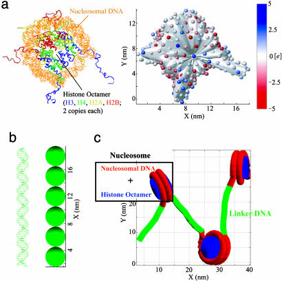
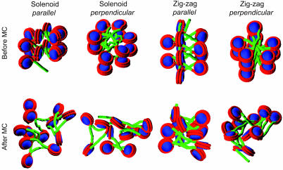
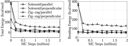
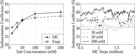
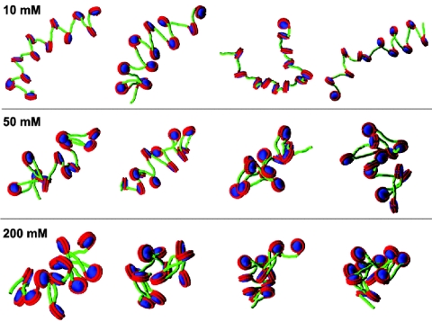
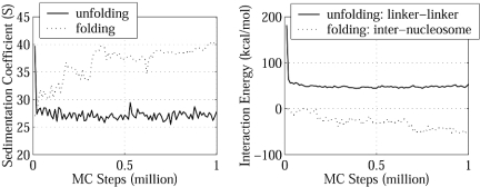
Although numerous experiments indicate that the chromatin fiber displays salt-dependent conformations, the associated molecular mechanism remains unclear. Here, we apply an irregular Discrete Surface Charge Optimization (DiSCO) model of the nucleosome with all histone tails incorporated to describe by Monte Carlo simulations salt-dependent rearrangements of a nucleosomal array with 12 nucleosomes. The ensemble of nucleosomal array conformations display salt-dependent condensation in good agreement with hydrodynamic measurements and suggest that the array adopts highly irregular 3D zig-zag conformations at high (physiological) salt concentrations and transitions into the extended "beads-on-a-string" conformation at low salt. Energy analyses indicate that the repulsion among linker DNA leads to this extended form, whereas internucleosome attraction drives the folding at high salt. The balance between these two contributions determines the salt-dependent condensation. Importantly, the internucleosome and linker DNA-nucleosome attractions require histone tails; we find that the H3 tails, in particular, are crucial for stabilizing the moderately folded fiber at physiological monovalent salt.


Figures

Fig. 1.
Fig. 1.
Models of nucleosome and linker DNA. (a Left) Crystal structure of nucleosome with complete modeled histone tails (PDB ID code 1KX5). (a Right) DiSCO model of the nucleosome with 300 effective charges located on an irregular surface. The surface was reduced by5Åto show the positions of the charges. The color scale indicates the value of each charge in the unit of e. (b Left) Linker DNA that connects two nucleosomes. (b Right) DNA bead model with six beads of 3-nm diameter. (c) Simplified representation of a short nucleosomal array showing how linker DNA connects nucleosomes.
Fig. 2.
Fig. 2.
Structures of nucleosomal arrays before and after MC simulations from different initial models. The initial models are constructed with nucleosome disk planes either parallel or perpendicular to the array helix axis before 2 million steps (6 million steps for the solenoid model with parallel nucleosomes) of MC simulations.
Fig. 3.
Fig. 3.
Total and bending energies as a function of MC steps for different initial models at 200 mM. Four different initial structures, representing the solenoid and zig-zag models with both parallel and perpendicular nucleosome orientations, are shown in Fig. 2. Data points are collected every 5,000 MC steps but are plotted every 200,000 steps.
Fig. 4.
Fig. 4.
Sedimentation coefficients s20,w as a function of salt concentration and MC steps. The experimental values (Expt.) are from ref. . Calculation of s20,w uses Eq. 1; every 10000th sample is used for the mean and standard deviation calculation.
Fig. 5.
Fig. 5.
Snapshots of the nucleosomal array at different salt concentrations. From left to right, the snapshots are taken after 0.5 million, 1 million, 1.5 million, and 2 million steps of the MC simulations.
Fig. 6.
Fig. 6.
Unfolding and folding simulations of nucleosomal array. (Left) s20,w during the unfolding simulation at 10 mM and the folding simulation at 200 mM. (Right) Linker–linker interaction energy during the unfolding simulation at 10 mM and internucleosome interaction energy during the folding simulation at 200 mM.

References

    1. van Holde, K. E. (1989) Chromatin (Springer, New York).
    1. Wolffe, A. (1995) Chromatin: Structure and Function (Academic, London).
    1. Wolffe, A. P. & Guschin, D. (2000) J. Struct. Biol. 129, 102-122. - PubMed
    1. Felsenfeld, G. & Groudine, M. (2003) Nature 421, 448-453. - PubMed
    1. Bednar, J., Horowitz, R. A., Grigoryev, S. A., Carruthers, L. M., Hansen, J. C., Koster, A. J. & Woodcock, C. L. (1998) Proc. Natl. Acad. Sci. USA 95, 14173-14178. - PMC - PubMed

Publication types

LinkOut - more resources