

Dendritic cell precursors are permissive to dengue virus and human immunodeficiency virus infection
- PMID: 15919883
- PMCID: PMC1143643
- DOI: 10.1128/JVI.79.12.7291-7299.2005
Dendritic cell precursors are permissive to dengue virus and human immunodeficiency virus infection
Abstract
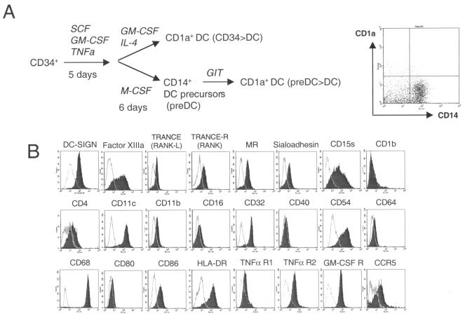
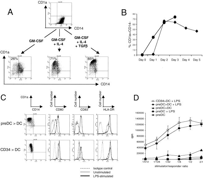
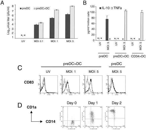
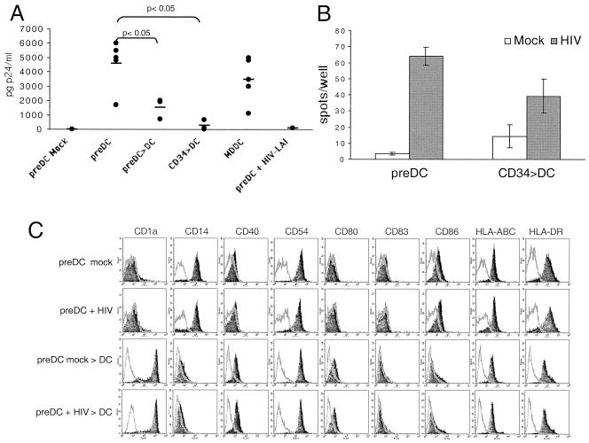
CD14(+) interstitial cells reside beneath the epidermis of skin and mucosal tissue and may therefore play an important role in viral infections and the shaping of an antiviral immune response. However, in contrast to dendritic cells (DC) or blood monocytes, these antigen-presenting cells (APC) have not been well studied. We have previously described long-lived CD14(+) cells generated from CD34(+) hematopoietic progenitors, which may represent model cells for interstitial CD14(+) APC. Here, we show that these cells carry DC-SIGN and differentiate into immature DC in the presence of granulocyte-macrophage colony-stimulating factor. We have compared the CD14(+) cells and the DC derived from these cells with respect to dengue virus and human immunodeficiency virus type 1 (HIV-1) infection. Both cell types are permissive to dengue virus infection, but the CD14(+) cells secrete the anti-inflammatory cytokine interleukin 10 and no tumor necrosis factor alpha. Regarding HIV, the CD14(+) cells are permissive to HIV-1, release higher p24 levels than the derived DC, and more efficiently activate HIV Pol-specific CD8(+) memory T cells. The CD14(+) DC precursors infected with either virus retain their DC differentiation potential. The results suggest that interstitial CD14(+) APC may contribute to HIV-1 and dengue virus infection and the shaping of an antiviral immune response.
Figures
Similar articles
-
CD34+ cell-derived CD14+ precursor cells develop into Langerhans cells in a TGF-beta 1-dependent manner.J Immunol. 1999 Nov 1;163(9):4869-77. J Immunol. 1999. PMID: 10528188
-
Differential infection of CD34+ cell-derived dendritic cells and monocytes with lymphocyte-tropic and monocyte-tropic HIV-1 strains.J Immunol. 1997 May 15;158(10):5035-42. J Immunol. 1997. PMID: 9144524
-
Mixed Langerhans cell and interstitial/dermal dendritic cell subsets emanating from monocytes in Th2-mediated inflammatory conditions respond differently to proinflammatory stimuli.J Leukoc Biol. 2006 Jul;80(1):45-58. doi: 10.1189/jlb.0205109. Epub 2006 Apr 13. J Leukoc Biol. 2006. PMID: 16614258
-
Monocytes, dendritic cells, and Langerhans cells in human immunodeficiency virus infection.Dermatol Clin. 1991 Jul;9(3):415-28. Dermatol Clin. 1991. PMID: 1873923 Review.
-
Mucosal dendritic cells and immunodeficiency viruses.J Infect Dis. 1999 May;179 Suppl 3:S427-30. doi: 10.1086/314798. J Infect Dis. 1999. PMID: 10099112 Review.
Cited by
-
Dengue virus serotype 2 blocks extracellular signal-regulated kinase and nuclear factor-κB activation to downregulate cytokine production.PLoS One. 2012;7(8):e41635. doi: 10.1371/journal.pone.0041635. Epub 2012 Aug 22. PLoS One. 2012. PMID: 22927911 Free PMC article.
-
Dengue fever in humanized NOD/SCID mice.J Virol. 2005 Nov;79(21):13797-9. doi: 10.1128/JVI.79.21.13797-13799.2005. J Virol. 2005. PMID: 16227299 Free PMC article.
-
Potential biomarkers for the clinical prognosis of severe dengue.Mem Inst Oswaldo Cruz. 2013 Sep;108(6):755-62. doi: 10.1590/0074-0276108062013012. Mem Inst Oswaldo Cruz. 2013. PMID: 24037198 Free PMC article.
-
Higher infection of dengue virus serotype 2 in human monocytes of patients with G6PD deficiency.PLoS One. 2008 Feb 13;3(2):e1557. doi: 10.1371/journal.pone.0001557. PLoS One. 2008. PMID: 18270558 Free PMC article.
-
Influence of dendritic cells on viral pathogenicity.PLoS Pathog. 2009 Jul;5(7):e1000384. doi: 10.1371/journal.ppat.1000384. Epub 2009 Jul 31. PLoS Pathog. 2009. PMID: 19649323 Free PMC article. Review.
References
-
- Anderson, D. M., E. Maraskovsky, W. L. Billingsley, W. C. Dougall, M. E. Tometsko, E. R. Roux, M. C. Teepe, R. F. DuBose, D. Cosman, and L. Galibert. 1997. A homologue of the TNF receptor and its ligand enhance T-cell growth and dendritic-cell function. Nature 390:175-179. - PubMed
-
- Caux, C., C. Dezutter-Dambuyant, D. Schmitt, and J. Banchereau. 1992. GM-CSF and TNF-α cooperate in the generation of dendritic Langerhans cells. Nature 360:258-261. - PubMed
-
- Caux, C., C. Massacrier, B. Dubois, J. Valladeau, C. Dezutter-Dambuyant, I. Durand, D. Schmitt, and S. Saeland. 1999. Respective involvement of TGF-beta and IL-4 in the development of Langerhans cells and non-Langerhans dendritic cells from CD34+ progenitors. J. Leukoc. Biol. 66:781-791. - PubMed
-
- Caux, C., B. Vanbervliet, C. Massacrier, C. Dezutter-Dambuyant, B. de Saint-Vis, C. Jacquet, K. Yoneda, S. Imamura, D. Schmitt, and J. Banchereau. 1996. CD34+ hematopoietic progenitors from human cord blood differentiate along two independent dendritic cell pathways in response to GM-CSF+TNFα. J. Exp. Med. 184:695-706. - PMC - PubMed
-
- Cerio, R., J. Spaull, G. F. Oliver, and W. E. Jones. 1990. A study of factor XIIIa and MAC 387 immunolabeling in normal and pathological skin. Am. J. Dermatopathol. 12:221-233. - PubMed
Publication types
MeSH terms
Substances
LinkOut - more resources
Full Text Sources
Medical
Research Materials
