. 2005 Jun;79(12):7478-91.
doi: 10.1128/JVI.79.12.7478-7491.2005.

Adenovirus binding to blood factors results in liver cell infection and hepatotoxicity

Affiliations

Adenovirus binding to blood factors results in liver cell infection and hepatotoxicity

Dmitry M Shayakhmetov et al. J Virol. 2005 Jun.

Abstract

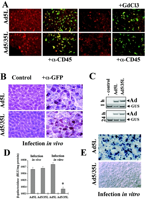
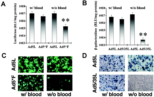
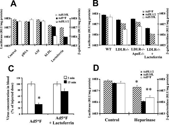
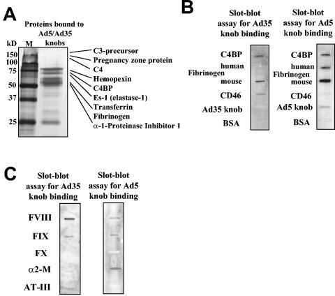
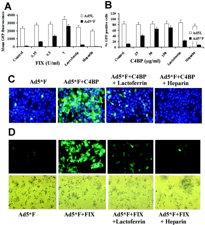
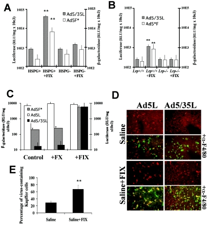
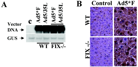
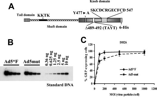
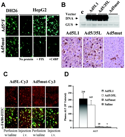
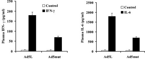
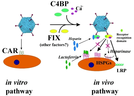
Adenoviruses (Ad) are efficient vehicles for gene delivery in vitro and in vivo. Therefore, they are a promising tool in gene therapy, particularly in the treatment of cancer and cardiovascular diseases. However, preclinical and clinical studies undertaken during the last decade have revealed a series of problems that limit both the safety and efficacy of Ad vectors, specifically after intravenous application. Major obstacles to clinical use include innate toxicity and Ad sequestration by nontarget tissues. The factors and mechanisms underlying these processes are poorly understood. The majority of intravenously injected Ad particles are sequestered by the liver, which in turn causes an inflammatory response characterized by acute transaminitis and vascular damage. Here, we describe a novel pathway that is used by Ad for infection of hepatocytes and Kupffer cells upon intravenous virus application in mice. We found that blood factors play a major role in targeting Ad vectors to hepatic cells. We demonstrated that coagulation factor IX and complement component C4-binding protein can bind the Ad fiber knob domain and provide a bridge for virus uptake through cell surface heparan sulfate proteoglycans and low-density lipoprotein receptor-related protein. An Ad vector, Ad5mut, which contained mutations in the fiber knob domain ablating blood factor binding, demonstrated significantly reduced infection of liver cells and liver toxicity in vivo. This study contributes to a better understanding of adenovirus-host interactions for intravenously applied vectors. It also provides a rationale for novel strategies to target adenovirus vector to specific tissues and to reduce virus-associated toxicity after systemic application.


Figures

FIG. 1.
FIG. 1.
Liver cell transduction with Ads in vivo and in vitro. Indicated Ads were injected into the tail vein of C57BL/6 mice. Livers of mice injected with Cy3-labeled Ad5L and Ad5/35L vectors were recovered 1 h (A) and 3 days (B) after virus administration. (A) Frozen liver sections either remained unstained or were stained with anti-mouse CD45 antibody (green) to detect Kupffer cells. Cy-3-labeled Ad particles appear in red. In settings with GdCl3, two doses of the drug were injected into mice 30 h and 6 h before Ad administration as described earlier (16). Note a significant reduction in virus accumulation within Kupffer cells after treatment of mice with GdCl3 (right). GdCl3 functionally inactivates Kupffer cells. (B) Paraffin sections of mouse livers 3 days after Ad virus administration were stained with hematoxilin-eosin (left) or stained with anti-GFP monoclonal antibody (brown). Kupffer cells are indicated by arrows. Note that GFP expression was detected in hepatocytes but not in Kupffer cells. (C) Southern blot analysis of Ad genomes in liver tissue 1 h and 24 h post virus administration. Mice were injected with saline (control) or Ad5L or Ad5/35L vectors. At the indicated time points, livers were recovered, and total DNA was purified and subjected to Southern blot analysis. For quantitative assessment of the data, equivalent loads of genomic DNA were confirmed by hybridization of the membranes with a probe specific for the mouse β-glucuronidase gene (GUS). After exposure to X-ray film, membranes were stripped and rehybridized with an Ad-specific probe to determine the levels of vector DNA associated with liver cells (Ad). (D) β-Galactosidase reporter gene activity in liver tissue (“Infection in vivo” bars), following intravenous administration of indicated Ad vectors or in purified mouse hepatocytes infected with Ad in vitro (“Infection in vitro” bars). (E) Purified mouse hepatocytes were infected with indicated Ad vectors and 48 h later stained in situ for β-galactosidase activity. Representative fields are shown. Studies shown in panels B through E were done with mice without GdCl3 treatment.
FIG. 2.
FIG. 2.
The role of blood factors in Ad transduction of hepatocytes in vivo. (A) Ad5L1 and Ad5*F express GFP and luciferase as reporters. (B) Ad5L2 and Ad5/35L express β-galactosidase (β-Gal) as a reporter. In the with-blood setting, Ad5L1/2, Ad5/35L, or Ad5*F was injected into the portal vein through a permanently placed catheter. Fifteen minutes later, hepatocytes were isolated for culture and analysis of reporter gene expression. In without-blood settings, the portal vein and vena cava inferior were canulated and blood was flushed from the liver through the portal vein. Virus was infused through the portal vein and the circulation between vena porta and vena cava was closed, allowing asanguinous isolated liver perfusion with virus-containing saline. Thirty minutes after virus application, hepatocyes were isolated by collagenase perfusion. In both settings, reporter gene expression in plated hepatocytes was analyzed 48 h postinfection. Four mice per group were analyzed for Ad5L1, Ad5*F, Ad5L2 and Ad5/35L variants with blood and for Ad5L1 without blood. Five mice per group were analyzed for Ad5L2 variant without blood, and seven and eight mice per group were analyzed for Ad5*F and Ad5/35L variants without blood, respectively. **, P < 0.001. There were no statistically significant differences between other paired variants. (C) Hepatocyte transduction with GFP-expressing Ad viruses (UV fluorescence). (D) Hepatocyte transduction with β-galactosidase-expressing Ad viruses after histochemical staining with X-galactosidase.
FIG. 3.
FIG. 3.
Analysis of hepatocellular receptors mediating the uptake of complexes between Ad and blood factors. (A) Competition of Ad infection with different ligands to hepatocellular receptors. The indicated competitors or saline (as a control) were injected into the portal vein through a catheter 5 min before virus administration. Hepatocytes were purified and reporter gene activities were analyzed as described in Materials and Methods. Three mice per group were analyzed for control, pBSA, asialofetuin (ASF), and hLDL groups, and five mice were analyzed for the lactoferrin group. *, P = 0.0056 for the hLDL Ad5L1/2 group, compared to control. For the other viruses in the hLDL group and all viruses in the lactoferrin group, there was a significant difference in hepatocyte transduction compared to the control group (P < 0.001). (B) Analysis of hepatocyte transduction in wild-type, ldlr−/−, and ldlr/ApoE−/− knockout mice. A total of 1011 viral particles were injected into the portal vein. After 15 min, hepatocytes were harvested, and reporter gene activities were analyzed 48 h later. (C) Blood persistence of Ad5*F in blood with or without lactoferrin administration. Lactoferrin was injected into the portal vein 5 min prior virus administration. * P < 0.01 for four samples. (D) Heparinase I was administrated into the tail vein of C57BL/6 mice 30 min before virus injection. Fifteen minutes after virus application, hepatocytes were harvested, plated, and analyzed for reporter gene activities 2 days postinfection. Three mice per group were analyzed for all control groups, four mice per group were analyzed for Ad5L1/2, and five mice per group were analyzed for Ad5F* and Ad5/35L (heparinase treated) groups. *, P < 0.01; **, P < 0.001.
FIG. 4.
FIG. 4.
Analysis of plasma proteins interacting with Ad fiber knob domains by mass spectrometry and protein slot blot assay. (A) Silver-stained polyacrylamide gel of proteins precipitated from mouse plasma with Ad5 and Ad35 fiber knob domains. (B and C) Slot blot analysis of direct binding of Ad fiber knob domains to purified plasma proteins (10 μg/lane) immobilized on nitrocellulose membrane. Human CD46 protein was used as a positive control to detect specific Ad35 fiber knob binding. Ad5, Ad35, and BSA were used to determine the levels of nonspecific binding. FVIII, FIX, and FX, coagulation factors VIII, IX and X, respectively; α2-M, α2-macroglobulin; AT-III, antithrombin III.
FIG. 5.
FIG. 5.
Interaction of Ad with C4BP and coagulation FIX mediates CAR-independent infection of human cells in vitro. (A) Dose-dependent transduction of human hepatoma HepG2 cells by Ad vectors with or without FIX (in units per milliliter, with 1 U corresponding to the physiological concentration of FIX in plasma). For infection inhibition experiments, 0.5 mg/ml of lactoferrin and 10 U/ml of heparin were used, with four mice per group analyzed. (B) Dose-dependent transduction of human hepatoma HepG2 cells by Ad5*F with or without C4BP (in micrograms per milliliter, where 150 μg/ml corresponds to the physiological concentration of C4BP in plasma), with four mice per group analyzed. (C) Expression of the GFP gene in human hepatoma HepG2 cells infected with Ad5*F in the presence of C4BP and different competitors 24 h post virus infection. Infections were done as described for panel B and in Materials and Methods. Cell nuclei were counterstained with DAPI (4-,6-diamidino-2-phenylindole) (blue). Representative fields with similar cell densities are shown. (D) Expression of GFP in primary human hepatocytes infected with Ad5*F in the presence of FIX and different competitors 24 h post virus infection. Infections were done as described for panel A and in Materials and Methods. Representative fields are shown. Upper panels demonstrate cells illuminated by UV light; lower panels demonstrate the same areas in visible light.
FIG. 6.
FIG. 6.
Interaction of Ad with coagulation FIX mediates CAR-independent infection of mouse cells in vitro and ex vivo. (A) Transduction of HSPG-expressing (CHO-K1) and HSPG-negative (CHO-pgsA745) cells by Ad5F* or Ad5/35L with or without FIX. (Averages and standard deviations for four independent experiments are shown.) **, P < 0.01. (B) Infection of mouse embryonic fibroblast MEF-1 (lrp+/+) or MEF-2 (lrp−/−) cells by Ad5F* or Ad5/35L with or without FIX. (C) Isolated, asanguinous liver perfusion of C57BL/6 mice with virus or saline plus human FX or FIX (1 U/ml). After virus perfusion, hepatocytes were isolated, and transgene expression was analyzed, with four mice analyzed for each virus group. **, P < 0.001. (D) Accumulation of Cy3-labeled Ad5L and Ad5/35L viruses (red) in F4/80-positive Kupffer cells (green) on liver sections 30 min after isolated liver perfusion with virus-containing saline with and without the addition of FIX. Note the absence of both Ad5L and Ad5/35L virus association with Kupffer cells when the livers were perfused in the absence of FIX. Representative fields of livers are shown, with three samples analyzed. (E) Quantitative representation of Ad accumulation in Kuppfer cells in mouse livers perfused with or without addition of FIX. Quantifications of virus-containing Kupffer cells on liver sections were conducted in a blinded fashion by independent experienced pathologists. At least 10 liver sections per experimental group were utilized to collect the data for statistical analysis. **, P < 0.01.
FIG. 7.
FIG. 7.
Accumulation of Ad genomic DNA in liver tissue and transduction of hepatocytes after vector administration into wild-type and FIX knockout mice. (A) Analysis of Ad5*F genomic DNA accumulation in liver tissue of wild-type or FIX knockout mice 72 h post vector administration, as determined by quantitative Southern blotting as shown in Fig. 1C. (B) Histological analysis of hepatocyte transduction after intravenous administration of Ad5*F vector to wild-type or FIX knockout mice. All procedures were done as described in Materials and Methods and in the legend to Fig. 1B.
FIG. 8.
FIG. 8.
Fiber structure of Ad mutated for binding to CAR and blood factors and its biological activity. (A) Schematic representations of mutations introduced into the Ad5L fiber knob domain ablating its binding to both CAR and blood factors. (B) Quantitative Southern blot analysis of purified Ad5*F and Ad5mut vector stocks. Virus DNA purified from 10 μl of vector stock was applied (in twofold serial dilutions) on an agarose gel together with serial dilutions of standard DNA (linearized adenovirus genome-containing plasmid). After hybridization with a 32P-labeled Ad-specific DNA probe and quantitation of signals with a phosphorimager, the concentrations of the indicated viruses were calculated. The concentration for Ad5*F was 8.6 × 10¹² virus particles per ml and the concentration for Ad5mut was 6.8 × 10¹² virus particles per ml. (C) Transduction of 293-DH26 cells with Ad5*F and Ad5mut vectors. (Averages and standard deviations for three independent experiments are shown.) For all further studies, an MOI of 1,000 virus particles per cell was used.
FIG. 9.
FIG. 9.
FIX-mediated infection of primary human hepatocytes and reduced infection of mouse liver with mutated Ad vector in vivo. (A) Visualization of GFP expression in 293-DH26 and HepG2 cells infected with Ad5*F and Ad5mut in the presence of absence of C4BP (150 μg/ml) and FIX (1 U/ml), with four mice analyzed per group. Representative fields with comparable cell density are shown. (B) Transduction of liver cells with Ad5L1, Ad5/35L-GFP, and Ad5mut vectors. Southern blot analysis (top) of vector DNA in mouse livers 72 h after intravenous administration of the indicated vectors. Control, liver DNA of a mouse injected with saline; GUS, single-copy mouse β-glucuronidase gene. GFP immunohistochemistry (bottom) of formalin-fixed and paraffin-embedded sections of livers harvested from mice injected with indicated viruses. (C) Detection of Cy3-labeled Ad5L1 and Ad5mut viruses (red) and F4/80-positive Kupffer cells (green) on liver sections 1 h after isolated liver perfusion with virus-containing saline with and without addition of FIX or after injection or viruses intravenously. Representative fields of livers are shown, with three mice analyzed per group. (D) Plasma ALT levels 72 h after tail vein injection of 1011 virus particles of the indicated Ad vectors or saline, with five mice analyzed per group.
FIG. 10.
FIG. 10.
Plasma levels of IFN-γ and IL-6 cytokines after administration of mice with Ad vectors. Mice were administered Ad5L or Ad5mut vectors; 6 h later, plasma samples were collected, and the levels of IFN-γ and IL-6 were analyzed by cytometric bead array as described in Materials and Methods (four mice per group).
FIG. 11.
FIG. 11.
Schematic representation of CAR-dependent and blood factor-dependent pathways of Ad infection and action of competitors (lactoferrin, heparin, and heparinase).

References

    1. Agnello, V., G. Abel, M. Elfahal, G. B. Knight, and Q. X. Zhang. 1999. Hepatitis C virus and other flaviviridae viruses enter cells via low density lipoprotein receptor. Proc. Natl. Acad. Sci. USA 96:12766-12771. - PMC - PubMed
    1. Alemany, R., and D. T. Curiel. 2001. CAR-binding ablation does not change biodistribution and toxicity of adenoviral vectors. Gene Ther. 8:1347-1353. - PubMed
    1. Ben-Gary, H., R. L. McKinney, T. Rosengart, M. L. Lesser, and R. G. Crystal. 2002. Systemic interleukin-6 responses following administration of adenovirus gene transfer vectors to humans by different routes. Mol. Ther. 6:287-297. - PubMed
    1. Bernt, K. M., D. S. Steinwaerder, S. Ni, Z. Y. Li, S. R. Roffler, and A. Lieber. 2002. Enzyme-activated prodrug therapy enhances tumor-specific replication of adenovirus vectors. Cancer Res. 62:6089-6098. - PubMed
    1. Branchereau, S., D. Calise, and N. Ferry. 1994. Factors influencing retroviral-mediated gene transfer into hepatocytes in vivo. Hum. Gene Ther. 5:803-808. - PubMed

Publication types

LinkOut - more resources