

Invasion of the central nervous system in a porcine host by nipah virus
- PMID: 15919907
- PMCID: PMC1143674
- DOI: 10.1128/JVI.79.12.7528-7534.2005
Invasion of the central nervous system in a porcine host by nipah virus
Abstract
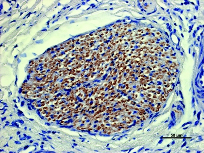
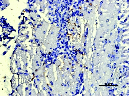
Nipah virus, a newly emerged zoonotic paramyxovirus, infects a number of species. Human infections were linked to direct contact with pigs, specifically with their body fluids. Clinical signs in human cases indicated primarily involvement of the central nervous system, while in pigs the respiratory system was considered the primary virus target, with only rare involvement of the central nervous system. Eleven 5-week-old piglets were infected intranasally, orally, and ocularly with 2.5 x 10(5) PFU of Nipah virus per animal and euthanized between 3 and 8 days postinoculation. Nipah virus caused neurological signs in two out of eleven inoculated pigs. The rest of the pigs remained clinically healthy. Virus was detected in the respiratory system (turbinates, nasopharynx, trachea, bronchus, and lung in titers up to 10(5.3) PFU/g) and in the lymphoreticular system (endothelial cells of blood and lymphatic vessels, submandibular and bronchiolar lymph nodes, tonsil, and spleen with titers up to 10(6) PFU/g). Virus presence was confirmed in the nervous system of both sick and apparently healthy animals (cranial nerves, trigeminal ganglion, brain, and cerebrospinal fluid, with titers up to 10(7.7) PFU/g of tissue). Nipah virus distribution was confirmed by immunohistochemistry. The study presents novel findings indicating that Nipah virus invaded the central nervous system of the porcine host via cranial nerves as well as by crossing the blood-brain barrier after initial virus replication in the upper respiratory tract.
Figures


Similar articles
-
Identifying Early Target Cells of Nipah Virus Infection in Syrian Hamsters.PLoS Negl Trop Dis. 2016 Nov 3;10(11):e0005120. doi: 10.1371/journal.pntd.0005120. eCollection 2016 Nov. PLoS Negl Trop Dis. 2016. PMID: 27812087 Free PMC article.
-
Bacterial infections in pigs experimentally infected with Nipah virus.Transbound Emerg Dis. 2008 May;55(3-4):165-74. doi: 10.1111/j.1865-1682.2008.01021.x. Transbound Emerg Dis. 2008. PMID: 18405339
-
Experimental Nipah virus infection in pigs and cats.J Comp Pathol. 2002 Feb-Apr;126(2-3):124-36. doi: 10.1053/jcpa.2001.0532. J Comp Pathol. 2002. PMID: 11945001
-
Pathogenesis of Hendra and Nipah virus infection in humans.J Infect Dev Ctries. 2013 Apr 17;7(4):308-11. doi: 10.3855/jidc.3648. J Infect Dev Ctries. 2013. PMID: 23592639 Review.
-
Henipaviruses in their natural animal hosts.Curr Top Microbiol Immunol. 2012;359:105-21. doi: 10.1007/82_2012_210. Curr Top Microbiol Immunol. 2012. PMID: 22476529 Review.
Cited by
-
Immunization strategies against henipaviruses.Curr Top Microbiol Immunol. 2012;359:197-223. doi: 10.1007/82_2012_213. Curr Top Microbiol Immunol. 2012. PMID: 22481140 Free PMC article. Review.
-
Nipah virus infects specific subsets of porcine peripheral blood mononuclear cells.PLoS One. 2012;7(1):e30855. doi: 10.1371/journal.pone.0030855. Epub 2012 Jan 27. PLoS One. 2012. PMID: 22303463 Free PMC article.
-
Identifying Early Target Cells of Nipah Virus Infection in Syrian Hamsters.PLoS Negl Trop Dis. 2016 Nov 3;10(11):e0005120. doi: 10.1371/journal.pntd.0005120. eCollection 2016 Nov. PLoS Negl Trop Dis. 2016. PMID: 27812087 Free PMC article.
-
Pathogenicity of Nipah henipavirus Bangladesh in a swine host.Sci Rep. 2019 Mar 26;9(1):5230. doi: 10.1038/s41598-019-40476-y. Sci Rep. 2019. PMID: 30914663 Free PMC article.
-
Henipaviruses: epidemiology, ecology, disease, and the development of vaccines and therapeutics.Clin Microbiol Rev. 2025 Mar 13;38(1):e0012823. doi: 10.1128/cmr.00128-23. Epub 2024 Dec 23. Clin Microbiol Rev. 2025. PMID: 39714175 Free PMC article. Review.
References
-
- Brown, C., D. J. King, and B. S. Seal. 1999. Pathogenesis of Newcastle disease in chickens experimentally infected with viruses of different virulence. Vet. Pathol. 36:125-132. - PubMed
-
- Carbone, K. M., and J. S. Wolinsky. 2001. Mumps virus, p. 1381-1400. In D. M. Knipe and P. M. Howley (ed.), Fields virology, 4th ed. Lippincott Williams and Wilkins, Philadelphia, Pa.
-
- Chua, K. B., W. J. Bellini, P. A. Rota, B. H. Harcourt, A. Tamin, S. K. Lam, T. G. Ksiazek, P. E. Rollin, S. R. Zaki, W.-J. Shieh, C. S. Goldsmith, D. J. Gubler, J. T. Roehring, B. Eaton, A. R. Gould, J. Olson, H. Field, P. Daniels, A. E. Ling, C. J. Peters, L. J. Anderson, and B. W. J. Mahy. 2000. Nipah virus: a recently emergent deadly paramyxovirus. Science 288:1432-1435. - PubMed
-
- Chua, K. B. 2003. Nipah virus outbreak in Malaysia. J. Clin. Virol. 26:265-275. - PubMed
Publication types
MeSH terms
LinkOut - more resources
Full Text Sources
Other Literature Sources
