. 2005 Jun;79(12):7609-16.
doi: 10.1128/JVI.79.12.7609-7616.2005.

Herpes simplex virus type 1 enters human epidermal keratinocytes, but not neurons, via a pH-dependent endocytic pathway

Affiliations

Herpes simplex virus type 1 enters human epidermal keratinocytes, but not neurons, via a pH-dependent endocytic pathway

Anthony V Nicola et al. J Virol. 2005 Jun.

Abstract

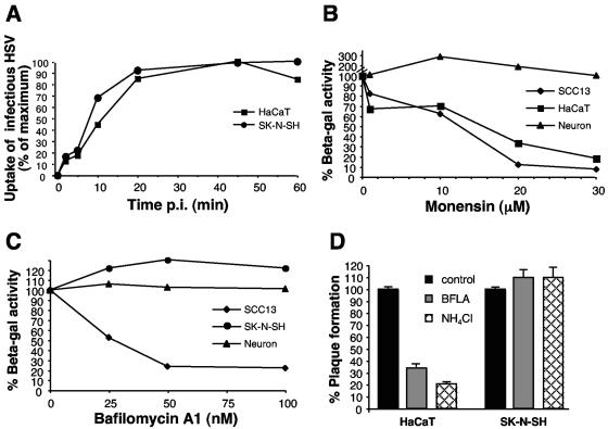
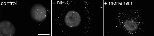
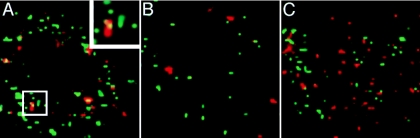
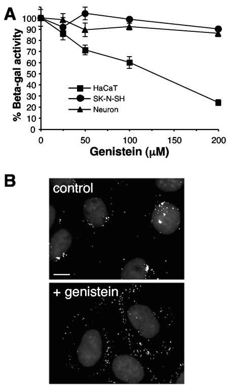
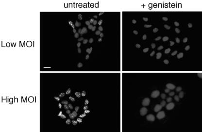
Herpes simplex virus (HSV) enters some laboratory cell lines via a pH-dependent, endocytic mechanism. We investigated whether this entry pathway is used in human cell types relevant to pathogenesis. Three different classes of lysosomotropic agents, which raise endosomal pH, blocked HSV entry into primary and transformed human keratinocytes, but not into human neurons or neuroblastoma lines. In keratinocytes, incoming HSV particles colocalized with markers of endocytic uptake. Treatment with the isoflavone genistein, an inhibitor of protein tyrosine kinases, reduced the delivery of incoming viral particles to the nuclear periphery and virus-induced gene expression in keratinocytes but not neurons. Moreover, in keratinocyte monolayer islets, HSV infected both the inner and outer cells in a genistein-sensitive manner, suggesting viral endocytosis from both basolateral and apical plasma membrane surfaces. Together, the results indicate that HSV enters human epidermal keratinocytes, but not neurons, by a low-pH, endocytic pathway that is dependent on host tyrosine phosphorylation. Thus, HSV utilizes fundamentally different cellular entry pathways to infect important target cell populations.


Figures

FIG. 1.
FIG. 1.
Effect of lysosomotropic agents on HSV infection of neurons and keratinocytes. (A) Uptake of infectious HSV from the cell surface. HSV-1 KOS K26GFP was bound to HaCaT keratinocytes or SK-N-SH neuroblastoma cells for 1 h at 4°C (MOI of 0.5). Cells were washed with PBS and incubated at 37°C, and extracellular virus was inactivated by acid treatment at the indicated times. At 8 hpi, cells were fixed and random fields of ∼1,000 cells in total were evaluated per sample. Cell number was determined by nuclear staining with DAPI, and infected, GFP-positive cells were counted. Maximum infectivity was set to 100%. (B, C) Effect of lysosomotropic agents on HSV-induced gene expression. Cells were pretreated with the indicated concentrations of agent for 1 h. Cells were infected with the lacZ+ HSV-1 strain KOS 7134 at an MOI of 1 for 7 h in the continued presence of agent. (D) Effect of lysosomotropic agents on HSV plaque formation. Cells were pretreated with 100 nM bafilomycin or 50 mM ammonium chloride for 1 h. HSV-1 KOS was added for 6 h at 37°C in the presence of the agent. The monolayers were washed and then incubated in normal medium for an additional 16 h. Plaques were visualized by immunoperoxidase staining with anti-HSV polyclonal sera. Each point represents the mean of quadruplicate wells.
FIG. 2.
FIG. 2.
Binding of HSV to the cell surface in the presence of various agents. HaCaT cells were treated for 1 h at 37°C with 30 μM monensin, 50 mM ammonium chloride, 100 nM bafilomycin A1 (BFLA), 200 μM genistein or 1 μg/ml heparin. 35S-labeled HSV-1 KOS was added (MOI of 3) in the continued presence of agents for 1 h at 4°C. Cells were washed with PBS, detergent lysates were prepared, and samples were analyzed with a liquid scintillation counter. Cell-associated cpm represents virus that was bound to the cell surface. The mean of three replicate samples ± standard error is shown.
FIG. 3.
FIG. 3.
Intracellular low pH is important for HSV entry into keratinocytes. Primary human keratinocytes were treated with 10 mM ammonium chloride, 5 μM monensin, or no agent for 1 h at 37°C. HSV-1 KOS K26GFP (MOI of 20) was then bound to the cells for 2 h at 4°C. Cells were washed with PBS, and then warmed medium containing the indicated inhibitors was added. Infection proceeded for 3 h in the presence of cycloheximide. Cells were washed with PBS and fixed in 3% paraformaldehyde. Nuclei were counterstained with DAPI. Punctate fluorescence indicates HSV particles. Cells were viewed with a ×63 oil immersion objective. The images are representative of the cell population. Bar, 10 μm.
FIG. 4.
FIG. 4.
Colocalization of incoming HSV with dextran, a marker of fluid-phase endocytosis. HaCaT (A, B) or SK-N-SH (C) cells were chilled for 15 min, and then HSV-1 KOS K26GFP was added for 2 h at 4°C. Cells were washed, and then warmed medium containing 1 mg/ml 70,000-kDa dextran-Texas red was added, and cells were rapidly warmed to 37°C. At 12 min (A, C) or 45 min (B) p.i., cells were fixed with 3% paraformaldehyde. A confocal slice of a single cell is shown for each. The inset in panel A is a magnification of the boxed area. Bar, 5 μm.
FIG. 5.
FIG. 5.
Role of host tyrosine phosphorylation in HSV entry and virus-induced gene expression. (A) HaCaT, fetal brain progenitor-derived neurons, or SK-N-SH cells were pretreated with the indicated concentrations of genistein for 1 h. Cells were infected with lacZ+ KOS 7134 at an MOI of 1 for 7 h in the continued presence of genistein. Entry was measured as the percentage of β-galactosidase activity relative to that obtained in the absence of genistein. Standard deviations are indicated. (B) Cells were treated for 1 h with 20 μM genistein (lower panel) or untreated (upper panel), and then HSV-1 KOS K26GFP (MOI of 20) was bound at 4°C for 2 h. Following a shift to 37°C for 2 h in the presence of genistein, cells were processed as described in the legend to Fig. 3. The images are representative of the cell population. Bar, 10 μm.
FIG. 6.
FIG. 6.
Effect of genistein on infection of keratinocyte islets. HaCaT cells were seeded at low density on glass coverslips. Islets formed by 3 to 4 days of culture. Cultures were treated with 50 μM genistein or mock treated for 1 h and were infected with HSV-1 KOS K26GFP at an MOI of 0.5 (low) or 5 (high) for 7 h in the presence or absence of genistein. Similar results were obtained with wortmannin (data not shown). Cultures were fixed with 3% paraformaldehyde, permeabilized, and then counterstained with DAPI. Newly synthesized VP26-GFP in the nucleus indicates successful entry and viral protein synthesis. Cells were viewed with a ×20 objective. Bar, 20 μm.

Similar articles

Cited by

References

    1. Berlin, R. D., and J. M. Oliver. 1980. Surface functions during mitosis. II. Quantitation of pinocytosis and kinetic characterization of the mitotic cycle with a new fluorescence technique. J. Cell Biol. 85:660-671. - PMC - PubMed
    1. Bodaghi, B., M. E. Slobbe-van Drunen, A. Topilko, E. Perret, R. C. Vossen, M. C. van Dam-Mieras, D. Zipeto, J. L. Virelizier, P. LeHoang, C. A. Bruggeman, and S. Michelson. 1999. Entry of human cytomegalovirus into retinal pigment epithelial and endothelial cells by endocytosis. Investig. Ophthalmol. Vis. Sci. 40:2598-2607. - PubMed
    1. Boukamp, P., R. T. Petrussevska, D. Breitkreutz, J. Hornung, A. Markham, and N. E. Fusenig. 1988. Normal keratinization in a spontaneously immortalized aneuploid human keratinocyte cell line. J. Cell Biol. 106:761-771. - PMC - PubMed
    1. Browne, H., B. Bruun, and T. Minson. 2001. Plasma membrane requirements for cell fusion induced by herpes simplex virus type 1 glycoproteins gB, gD, gH and gL. J. Gen. Virol. 82:1419-1422. - PubMed
    1. Cai, W., and P. A. Schaffer. 1991. A cellular function can enhance gene expression and plating efficiency of a mutant defective in the gene for ICP0, a transactivating protein of herpes simplex virus type 1. J. Virol. 65:4078-4090. - PMC - PubMed

LinkOut - more resources