. 2005 Jun;79(12):7617-28.
doi: 10.1128/JVI.79.12.7617-7628.2005.

Ikappabeta-related vankyrin genes in the Campoletis sonorensis ichnovirus: temporal and tissue-specific patterns of expression in parasitized Heliothis virescens lepidopteran hosts

Affiliations

Ikappabeta-related vankyrin genes in the Campoletis sonorensis ichnovirus: temporal and tissue-specific patterns of expression in parasitized Heliothis virescens lepidopteran hosts

Jeremy A Kroemer et al. J Virol. 2005 Jun.

Abstract

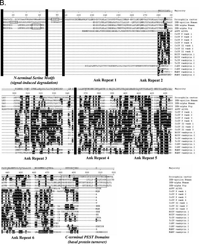
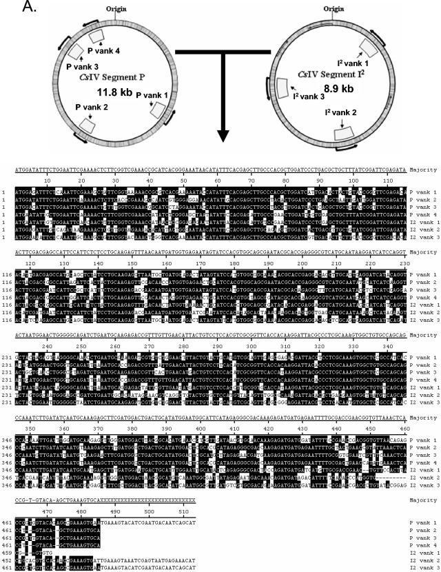
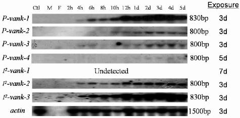
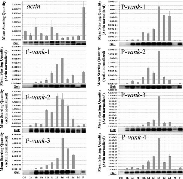
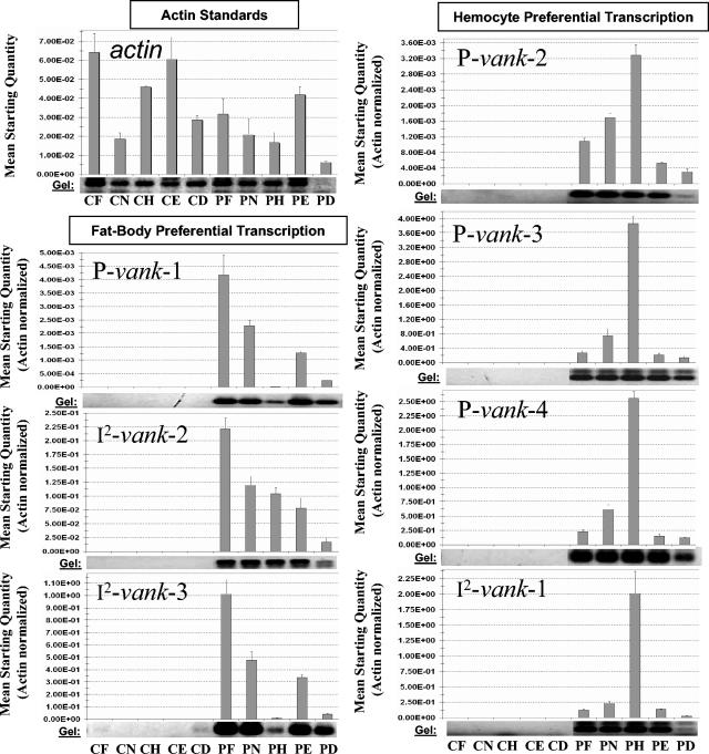
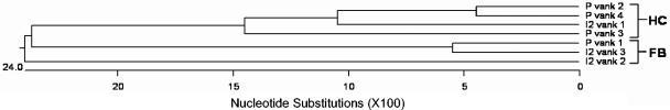
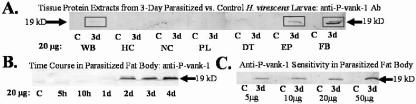
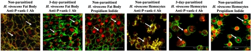
Polydnaviruses (PDVs) are unusual insect viruses that occur in obligate symbiotic associations with parasitic ichneumonid (ichnoviruses, or IVs) and braconid (bracoviruses, or BVs) wasps. PDVs are injected with eggs, ovarian proteins, and venom during parasitization. Following infection of cells in host tissues, viral genes are expressed and their products function to alter lepidopteran host physiology, enabling endoparasitoid development. Here we describe the Campoletis sonorensis IV viral ankyrin (vankyrin) gene family and its transcription. The seven members of this gene family possess ankyrin repeat domains that resemble the inhibitory domains of the Drosophila melanogaster NF-kappabeta transcription factor inhibitor (Ikappabeta) cactus. vankyrin gene expression is detected within 2 to 4 h postparasitization (p.p.) in Heliothis virescens hosts and reaches peak levels by 3 days p.p. Our data indicate that vankyrin genes from the C. sonorensis IV genome are differentially expressed in the tissues of parasitized hosts and can be divided into two subclasses: those that target the host fat body and those that target host hemocytes. Polyclonal antibodies raised against a fat-body targeting vankyrin detected a 19-kDa protein in crude extracts prepared from the 3 days p.p. fat body. Vankyrin-specific Abs localized to 3-day p.p. fat-body and hemocyte nuclei, suggesting a role for vankyrin proteins in the nuclei of C. sonorensis IV-infected cells. These data are evidence for divergent tissue specificities and targeting of multigene families in IVs. We hypothesize that PDV vankyrin genes may suppress NF-kappabeta activity during immune responses and developmental cascades in parasitized lepidopteran hosts of C. sonorensis.


Figures

FIG. 1.
FIG. 1.
A. Position of vankyrin (vank) genes on segments P and I2 of the C. sonorensis IV (CsIV) genome, and DNA sequence alignment of the seven gene family variants. Black residues indicate matches to the vankyrin consensus sequence. B. Protein sequence alignment of the C. sonorensis IV vankyrin gene family with members of the Iκβ gene family and representative vankyrin homologs from the HfIV, TrIV, MdBV, and CcBV PDV genomes. C. sonorensis IV vankyrin proteins align with ankyrin repeat domains other PDV vankyrins and with those of typical Iκβs. Black residues indicate matches to the consensus sequence. Locations of Drosophila cactus ankyrin repeats 1 to 6 have been denoted according to Dushay et al. (18). PDV vankyrin proteins lack N- and C-terminal destruction domains (black boxes) that are sensitive to protease activity and are involved in the regulation of cellular activity. Sequences removed to optimize the alignment are indicated by vertical black lines. Alignments were created with MEGALIGN software (DNASTAR, Madison, WI). NCBI accession numbers: Drosophila cactus (A44269), Iκβɛ human (O00221), Iκβα human (A39935), Iκβα pig (CAA84619), ASFV A238L (NP_042733), HfIV vankyrin 1(AAS90270), HfIV vankyrin 2 (AAX24120), TrIV vankyrin 1 and TrIV vankyrin 2 (AY940454), CcBV vankyrin 1 (CAG17493), CcBV vankyrin 2 (CAG17492), MdBV vankyrin 1 (AAW51804), MdBV vankyrin 2 (AAW51781).
FIG. 1.
FIG. 1.
A. Position of vankyrin (vank) genes on segments P and I2 of the C. sonorensis IV (CsIV) genome, and DNA sequence alignment of the seven gene family variants. Black residues indicate matches to the vankyrin consensus sequence. B. Protein sequence alignment of the C. sonorensis IV vankyrin gene family with members of the Iκβ gene family and representative vankyrin homologs from the HfIV, TrIV, MdBV, and CcBV PDV genomes. C. sonorensis IV vankyrin proteins align with ankyrin repeat domains other PDV vankyrins and with those of typical Iκβs. Black residues indicate matches to the consensus sequence. Locations of Drosophila cactus ankyrin repeats 1 to 6 have been denoted according to Dushay et al. (18). PDV vankyrin proteins lack N- and C-terminal destruction domains (black boxes) that are sensitive to protease activity and are involved in the regulation of cellular activity. Sequences removed to optimize the alignment are indicated by vertical black lines. Alignments were created with MEGALIGN software (DNASTAR, Madison, WI). NCBI accession numbers: Drosophila cactus (A44269), Iκβɛ human (O00221), Iκβα human (A39935), Iκβα pig (CAA84619), ASFV A238L (NP_042733), HfIV vankyrin 1(AAS90270), HfIV vankyrin 2 (AAX24120), TrIV vankyrin 1 and TrIV vankyrin 2 (AY940454), CcBV vankyrin 1 (CAG17493), CcBV vankyrin 2 (CAG17492), MdBV vankyrin 1 (AAW51804), MdBV vankyrin 2 (AAW51781).
FIG. 2.
FIG. 2.
Northern blots for H. virescens actin and six of the seven C. sonorensis IV vankyrin genes over the time course (2 h to 5 days) of C. sonorensis parasitization. Signals for individual vankyrin variants were detected after 3 to 5 days of exposure to X-ray film (right panel). An H. virescens actin probe verified that approximately equivalent amounts of total RNA were loaded in each well (∼30 μg/lane). Ctl, nonparasitized control H. virescens; M, male C. sonorensis;, F, female C. sonorensis; h, hours postparasitization; d, days postparasitization.
FIG. 3.
FIG. 3.
Relative quantitative real-time PCR for H. virescens actin and the seven C. sonorensis IV vankyrin genes over a 2-h to 6-day time course of parasitization. All values for viral genes were normalized to actin controls to account for variations in cDNA pools. All viral genes were expressed by 2 h p.p. and showed peak transcription by 3 days p.p. in H. virescens larvae. Error bars represent +1 standard deviations from the mean starting quantity calculated for each time point. Ctl, control H. virescens; h, hours p.p.; d, days p.p.; M, male C. sonorensis; F, female C. sonorensis. PCR amplimers were run on 1% agarose gels (photos) to verify the presence of DNA fragments of the correct size. No viral-specific amplimers were detected for cDNA pools of nonparasitized H. virescens control larvae.
FIG. 4.
FIG. 4.
Relative quantitative real-time PCR for H. virescens actin and the seven C. sonorensis IV vankyrin genes in selected tissues of control and 3-day-p.p. H. virescens larvae. All values for viral genes were normalized to actin controls to correct for variations between cDNA pools. Viral genes showed preferential targeting for transcription in different tissues of the parasitized insect. Expression of the P-vank-1, I2-vank-2, and I2-vank-3 viral genes was higher in the parasitized fat body relative to other infected tissues. The P-vank-2, P-vank-3, P-vank-4, and I2-vank-1 genes show highest levels of expression in the parasitized hemocytes. Error bars represent +1 standard deviation from the mean. CF, nonparasitized (np) fat body; CN, np nerve cord; CH, np hemocytes; CE, np epidermal layer (including trachea, muscles, and epidermis); CD, nonparasitized digestive tract (incuding gut and malpighian tubules); PF, 3-day-p.p. fat body; PN, 3-day-p.p. nerve cord; PH, 3-day-p.p. hemocytes; PE, 3-day-p.p. epidermal layer; PD, 3-day-p.p. digestive tract. PCR products for the viral genes were detected in all parasitized tissues examined (gels). No viral-specific amplimers were detected for cDNA pools of nonparasitized tissues.
FIG. 5.
FIG. 5.
Phylogenetic tree constructed via the Clustal W method of DNA alignment. The tree is rooted based upon assumption of the biological clock. HC, hemocyte-specific transcription; FB, fat body-specific transcription. Alignment parameters: gap penalty, 15; gap length penalty, 6.66; DNA weight matrix, International Union of Biochemistry. Tree was assembled with MEGALIGN software (DNASTAR, Madison, WI).
FIG. 6.
FIG. 6.
Western blots. A. Tissue specificity of the anti-P-vank-1 Ab in 3-day-p.p. insects. Crude protein extracts were prepared from nonparasitized control (C) and 3-day-parasitized (3-day) H. virescens whole-body tissue (WB), hemocytes (HC), nerve cord (NC), hemolymph plasma (PL), digestive tract (DT), epidermal layer (EP), and fat body tissue (FB). A 19-kDa protein of the predicted size from parasitized larval whole-body, epidermal layer, and fat-body extracts reacted specifically with the anti-P-vank-1 Ab. The signal appeared by 1 day p.p. in the fat body (B), and the Ab was sensitive to as little as 5 μg (C) of extract from the 3-day-p.p. fat body.
FIG. 7.
FIG. 7.
Immunofluorescence assays using anti-P-vank-1 and anti-P-vank-4 Abs. The anti-P-vank-1 (red) antibody localized to the 3-day-p.p. fat-body nucleus (arrows). Nonparasitized H. virescens fat-body tissue showed no specific nuclear staining (arrows). Propidium iodide staining of fat-body tissue verified the subcellular localization of cell nuclei (arrows). The anti-P-ank-4 antibody (red) localized to the 3-day-p.p. hemocyte nucleus (arrows). There was no specific staining in nonparasitized H. virescens hemocytes (arrows). Propidium iodide staining of hemocytes verified the subcellular localization of cell nuclei (arrows). All cells were counterstained with FITC-Phalloidin (green) to visualize F-actin. Magnification, ×100. Scale bar, 50 μm.

Similar articles

Cited by

References

    1. Andrade, M. A., C. Perez-Iratxeta, and C. P. Ponting. 2001. Protein repeats: structures, functions, and evolution. J. Struct. Biol. 134:117-131. - PubMed
    1. Asgari, S., M. Hellers, and O. Schmidt. 1996. Host haemocyte inactivation by an insect parasitoid: transient expression of a polydnavirus gene. J. Gen. Virol. 77:2653-2662. - PubMed
    1. Beck, M., and M. R. Strand. 2003. RNA interference silences Microplitis demolitor bracovirus genes and implicates glc1.8 in disruption of adhesion in infected host cells. Virology 314:521-535. - PubMed
    1. Beg, A. A., and A. S. Baldwin, Jr. 1993. The IκB proteins: multifunctional regulators of Rel/NF-κB transcription factors. Genes Dev. 7:2064-2070. - PubMed
    1. Begun, D. J., and P. Whitley. 2000. Adaptive evolution of relish, a Drosophila NF-κβ/Iκβ protein. Genetics 154:1231-1238. - PMC - PubMed

Publication types

MeSH terms

LinkOut - more resources