Murine coronavirus evolution in vivo: functional compensation of a detrimental amino acid substitution in the receptor binding domain of the spike glycoprotein
- PMID: 15919915
- PMCID: PMC1143675
- DOI: 10.1128/JVI.79.12.7629-7640.2005
Murine coronavirus evolution in vivo: functional compensation of a detrimental amino acid substitution in the receptor binding domain of the spike glycoprotein
Abstract
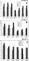
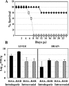
Murine coronavirus A59 strain causes mild to moderate hepatitis in mice. We have previously shown that mutants of A59, unable to induce hepatitis, may be selected by persistent infection of primary glial cells in vitro. These in vitro isolated mutants encoded two amino acids substitutions in the spike (S) gene: Q159L lies in the putative receptor binding domain of S, and H716D, within the cleavage signal of S. Here, we show that hepatotropic revertant variants may be selected from these in vitro isolated mutants (Q159L-H716D) by multiple passages in the mouse liver. One of these mutants, hr2, was chosen for more in-depth study based on a more hepatovirulent phenotype. The S gene of hr2 (Q159L-R654H-H716D-E1035D) differed from the in vitro isolates (Q159L-H716D) in only 2 amino acids (R654H and E1035D). Using targeted RNA recombination, we have constructed isogenic recombinant MHV-A59 viruses differing only in these specific amino acids in S (Q159L-R654H-H716D-E1035D). We demonstrate that specific amino acid substitutions within the spike gene of the hr2 isolate determine the ability of the virus to cause lethal hepatitis and replicate to significantly higher titers in the liver compared to wild-type A59. Our results provide compelling evidence of the ability of coronaviruses to rapidly evolve in vivo to highly virulent phenotypes by functional compensation of a detrimental amino acid substitution in the receptor binding domain of the spike glycoprotein.
Figures


Similar articles
-
Targeted recombination within the spike gene of murine coronavirus mouse hepatitis virus-A59: Q159 is a determinant of hepatotropism.J Virol. 1998 Dec;72(12):9628-36. doi: 10.1128/JVI.72.12.9628-9636.1998. J Virol. 1998. PMID: 9811696 Free PMC article.
-
Hepatitis mutants of mouse hepatitis virus strain A59.Adv Exp Med Biol. 1995;380:577-82. doi: 10.1007/978-1-4615-1899-0_92. Adv Exp Med Biol. 1995. PMID: 8830545
-
Altered pathogenesis of a mutant of the murine coronavirus MHV-A59 is associated with a Q159L amino acid substitution in the spike protein.Virology. 1997 Dec 8;239(1):1-10. doi: 10.1006/viro.1997.8877. Virology. 1997. PMID: 9426441 Free PMC article.
-
Receptor specificity and receptor-induced conformational changes in mouse hepatitis virus spike glycoprotein.Adv Exp Med Biol. 2001;494:173-81. doi: 10.1007/978-1-4615-1325-4_29. Adv Exp Med Biol. 2001. PMID: 11774465 Review. No abstract available.
-
Murine coronavirus neuropathogenesis: determinants of virulence.J Neurovirol. 2010 Nov;16(6):427-34. doi: 10.3109/13550284.2010.529238. Epub 2010 Nov 12. J Neurovirol. 2010. PMID: 21073281 Free PMC article. Review.
Cited by
-
Protective role of Toll-like Receptor 3-induced type I interferon in murine coronavirus infection of macrophages.Viruses. 2012 May;4(5):901-23. doi: 10.3390/v4050901. Epub 2012 May 24. Viruses. 2012. PMID: 22754655 Free PMC article.
-
Amino acid substitutions in the s2 region enhance severe acute respiratory syndrome coronavirus infectivity in rat angiotensin-converting enzyme 2-expressing cells.J Virol. 2007 Oct;81(19):10831-4. doi: 10.1128/JVI.01143-07. Epub 2007 Jul 25. J Virol. 2007. PMID: 17652383 Free PMC article.
-
Coronavirus pathogenesis.Adv Virus Res. 2011;81:85-164. doi: 10.1016/B978-0-12-385885-6.00009-2. Adv Virus Res. 2011. PMID: 22094080 Free PMC article. Review.
-
Antiviral effects of antisense morpholino oligomers in murine coronavirus infection models.J Virol. 2007 Jun;81(11):5637-48. doi: 10.1128/JVI.02360-06. Epub 2007 Mar 7. J Virol. 2007. PMID: 17344287 Free PMC article.
-
Homologous 2',5'-phosphodiesterases from disparate RNA viruses antagonize antiviral innate immunity.Proc Natl Acad Sci U S A. 2013 Aug 6;110(32):13114-9. doi: 10.1073/pnas.1306917110. Epub 2013 Jul 22. Proc Natl Acad Sci U S A. 2013. PMID: 23878220 Free PMC article.
References
Publication types
MeSH terms
Substances
Grants and funding
LinkOut - more resources
Full Text Sources

