. 2005 Jun;79(12):7648-57.
doi: 10.1128/JVI.79.12.7648-7657.2005.

Hepatitis C virus core protein suppresses NF-kappaB activation and cyclooxygenase-2 expression by direct interaction with IkappaB kinase beta

Affiliations

Hepatitis C virus core protein suppresses NF-kappaB activation and cyclooxygenase-2 expression by direct interaction with IkappaB kinase beta

Myungsoo Joo et al. J Virol. 2005 Jun.

Abstract

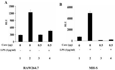
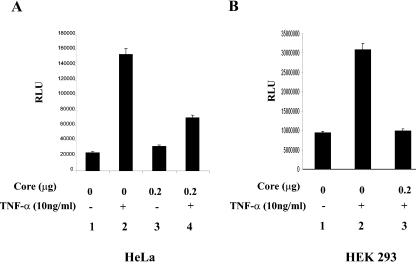
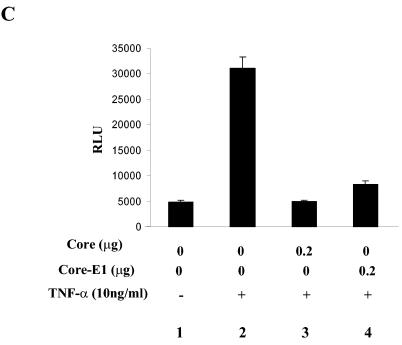
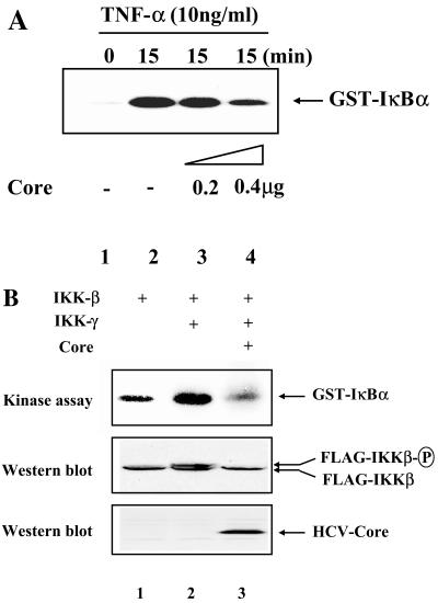
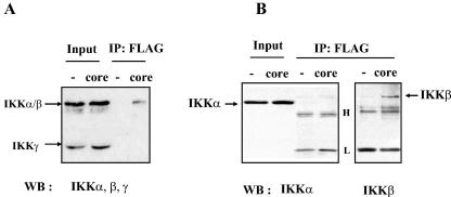
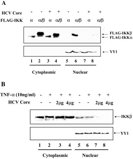
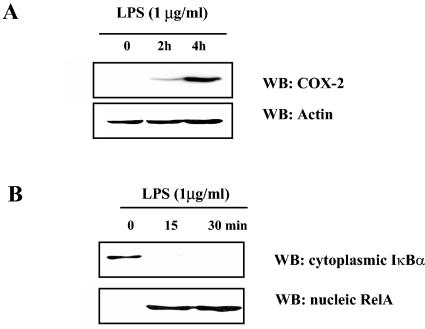
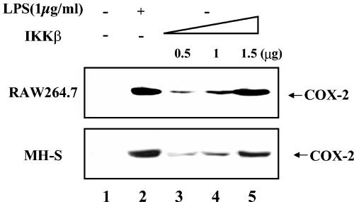
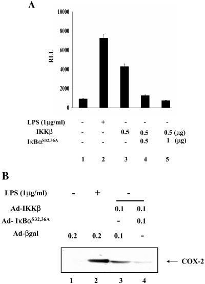
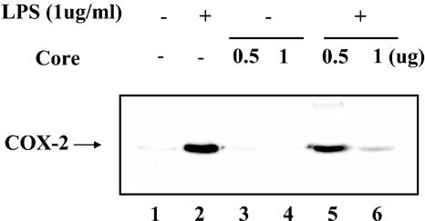
In addition to hepatocytes, hepatitis C virus (HCV) infects immune cells, including macrophages. However, little is known concerning the impact of HCV infection on cellular functions of these immune effector cells. Lipopolysaccharide (LPS) activates IkappaB kinase (IKK) signalsome and NF-kappaB, which leads to the expression of cyclooxygenase-2 (COX-2), which catalyzes production of prostaglandins, potent effectors on inflammation and possibly hepatitis. Here, we examined whether expression of HCV core interferes with IKK signalsome activity and COX-2 expression in activated macrophages. In reporter assays, HCV core inhibited NF-kappaB activation in RAW 264.7 and MH-S murine macrophage cell lines treated with bacterial LPS. HCV core inhibited IKK signalsome and IKKbeta kinase activities induced by tumor necrosis factor alpha in HeLa cells and coexpressed IKKgamma in 293 cells, respectively. HCV core was coprecipitated with IKappaKappabeta and prevented nuclear translocation of IKKbeta. NF-kappaB activation by either LPS or overexpression of IKKbeta was sufficient to induce robust expression of COX-2, which was markedly suppressed by ectopic expression of HCV core. Together, these data indicate that HCV core suppresses IKK signalsome activity, which blunts COX-2 expression in macrophages. Additional studies are necessary to determine whether interrupted COX-2 expression by HCV core contributes to HCV pathogenesis.


Figures

FIG. 1.
FIG. 1.
HCV core inhibits NF-κB activated by LPS. RAW 264.7 (A) and MH-S (B) cells were transfected with 0.5 μg of NF-κB-Luc and 0.2 μg of tk-Renilla-Luc along with the indicated amounts of the HCV core-expressing plasmid. The total amount of transfected plasmid was adjusted with an empty vector plasmid. NF-κB activity elicited by LPS was measured after treating cells with LPS for 4 h (lanes 1 and 2). To examine an effect of the HCV core on NF-κB-mediated transcriptional activity, the plasmid encoding HCV core was transfected without or with treatment with LPS (1 μg/ml) for 4 h (lanes 3 and 4).
FIG. 2.
FIG. 2.
HCV core inhibits NF-κB activated by TNF-α in other cell types. HeLa (A) and HEK 293 (B) cells were transfected with 0.2 μg of the NF-κB-Luc and 0.1 μg of the tk-Renilla-Luc reporter construct, along with the indicated amount of the HCV core-expressing plasmid. The total amount of transfected plasmid was adjusted with an empty vector plasmid. NF-κB-mediated transcriptional activity was measured after treating cells with TNF-α (10 ng/ml) for 6 h. (C) A possible effect of HCV E1 on HCV core was examined. HeLa cells were transfected with two reporter constructs, indicated above, along with either the plasmid encoding HCV core or a plasmid encoding HCV core and E1, and treated with TNF-α as described above. NF-κB-mediated transcription activity was measured by assaying luciferase activity. The result shown here is a representative of three independent experiments.
FIG. 2.
FIG. 2.
HCV core inhibits NF-κB activated by TNF-α in other cell types. HeLa (A) and HEK 293 (B) cells were transfected with 0.2 μg of the NF-κB-Luc and 0.1 μg of the tk-Renilla-Luc reporter construct, along with the indicated amount of the HCV core-expressing plasmid. The total amount of transfected plasmid was adjusted with an empty vector plasmid. NF-κB-mediated transcriptional activity was measured after treating cells with TNF-α (10 ng/ml) for 6 h. (C) A possible effect of HCV E1 on HCV core was examined. HeLa cells were transfected with two reporter constructs, indicated above, along with either the plasmid encoding HCV core or a plasmid encoding HCV core and E1, and treated with TNF-α as described above. NF-κB-mediated transcription activity was measured by assaying luciferase activity. The result shown here is a representative of three independent experiments.
FIG. 3.
FIG. 3.
The HCV core protein inhibits IKK kinase activity. (A) HeLa cells were transfected with either an empty vector (lanes 1 and 2) or increasing amounts of the HCV core-expressing plasmid (lanes 3 and 4). The total amount of transfected plasmid was normalized as indicated in Materials and Methods. At 48 h posttransfection, the cells were treated with TNF-α (10 ng/ml) for 15 min and the cytoplasmic fraction was prepared for immunoprecipitation of IKK with anti-IKKβ antibody. Immune complex, after being extensively washed, was incubated with GST-IκBα and [γ-32P]ATP to measure IKK kinase activity. (B) HEK 293 cells were transfected with 0.1 μg of the FLAG-tagged IKKβ-expressing plasmid along with 0.5 μg of a plasmid encoding IKKγ (lanes 2 and 3) and 0.5 μg of the HCV core-expressing plasmid (lane 3). At 48 h posttransfection, total cell lysate was prepared for immunoprecipitation with M2 antibody to capture IKKβ. Immune complexes were incubated with GST-IκBα and [γ-32P]ATP to measure IKKβ kinase activity (top panel). Expression levels of IKKβ and the HCV core were determined by Western blotting with M2 antibody and anti-HCV core antibody, respectively (middle and bottom panels).
FIG. 4.
FIG. 4.
The HCV core protein interacts with IKKβ. HeLa cells were transfected with either 2 μg of an empty vector plasmid or 1 μg of a plasmid encoding FLAG-tagged HCV core that was normalized with the empty vector plasmid to 2 μg. At 48 h posttransfection, cell lysate was prepared for immunoprecipitation of FLAG-tagged HCV core with M2 antibody. Immune complex captured by protein A-Sepharose was separated by SDS-PAGE and analyzed by Western blotting. (A) HCV core protein immunoprecipitated with IKK. Blot was incubated with anti-IKKα, -β, and -γ antibodies (rabbit polyclonal antibodies) to reveal subunits of IKK that interact with the HCV core protein. One-tenth of the total cell lysate used for immunoprecipitation was loaded as a positive control for IKKα, -β, and -γ. (B) The HCV core protein interacts with IKKβ. Total cell lysate was prepared from transfected cells and incubated with M2 antibody. One-tenth of the total cell lysate used for immunoprecipitation was loaded as a positive control for IKKα. Precipitated immune complex was analyzed by Western blotting after SDS-PAGE separation. The blot was incubated with either anti-IKKα antibody or anti-IKKβ (mouse monoclonal) antibody to reveal IKKα and IKKβ, respectively.
FIG. 5.
FIG. 5.
The HCV core protein interferes with localization of IKKβ to the nucleus. (A) The HCV core protein interferes with nuclear localization of transfected IKKβ. HEK 293 cells were transfected with 0.5 μg of the plasmid encoding FLAG-tagged IKKα and IKKβ with (lanes 3 and 4 and lanes 7 and 8) or without (lanes 1 and 2 and lanes 5 and 6) 2 μg of the plasmid encoding HCV core. The total amount of transfected plasmid was adjusted to 3 μg with an empty host plasmid. Cytoplasmic and nuclear fractions were prepared as described in Materials and Methods. Ten micrograms of cytoplasmic proteins and 50 μg of nuclear proteins were analyzed by SDS-PAGE and Western blotting with M2 antibody to reveal the location of IKKα and -β. To ensure equal loading of nuclear proteins, nuclear protein YY1 was Western blotted after stripping membrane. (B) The HCV core protein interferes with nuclear localization of endogenous IKKβ. HEK 293 cells were transfected with either 4 μg of an empty host plasmid (lanes 1 and 2 and lanes 5 and 6) or a different dose of the plasmid encoding HCV core (lanes 3 and 4 and 7 and 8). The total amount of transfected plasmid was adjusted to 4 μg with an empty host plasmid. At 48 h posttransfection, transfected cells were treated with TNF-α (10 ng/ml) for 30 min before preparing cytoplasmic and nuclear fractions. Fifty micrograms of each fraction was analyzed by Western blotting to reveal endogenous IKKβ. To ensure equal loading of nuclear protein, the membrane was stripped and reblotted to reveal nuclear protein YY1.
FIG. 6.
FIG. 6.
Casual relationship between NF-κB activation and COX-2 induction. (A) Murine macrophage cell line RAW 264.7 was treated with 1 μg/ml of LPS for the indicated times, and 20 μg of total cell lysate was analyzed for COX-2 expression. To ensure equal loading, membrane was stripped and Western blotted for actin. (B) After LPS treatment of RAW 264.7 cells for the indicated times, Western blotting was performed to reveal IκBα in the cytoplasm and RelA in the nucleus.
FIG. 7.
FIG. 7.
NF-κB activation confers COX-2 expression. RAW 264.7 and MH-S cells were transiently transfected with either 1.5 μg of an empty vector plasmid (lanes 1 and 2) or an increasing amount of a plasmid that encodes FLAG-tagged IKKβ (lanes 3 to 5). The total amount of transfected DNA was normalized as described in Materials and Methods. Transfected cells were further incubated without serum overnight and treated with or without LPS (1 μg/ml) for 4 h. Expression of COX-2 was measured by Western blotting with 20 μg of total cell lysate.
FIG. 8.
FIG. 8.
COX-2 induction by NF-κB is suppressed by the superrepressor IκBαS32,36A. (A) IκBαS32,36A inhibits NF-κB activated by IKKβ. RAW 264.7 cells were transfected with 0.2 μg of a tk-Renilla luciferase construct (tk-Renilla-Luc) and 0.5 μg of a luciferase reporter construct containing NF-κB-binding sites (NF-κB-Luc) along with an empty vector plasmid (lanes 1 and 2) or the indicated amounts of the IKKβ-expressing plasmid (lanes 3 to 5). To examine the inhibitory effect of IκBαS32,36A on NF-κB activated by IKKβ, an increasing amount of a plasmid encoding IκBαS32,36A was cotransfected (lanes 4 and 5). At 48 h posttransfection, the cell extract was prepared and enzymatic activity was measured. The result shown here is a representative of three independent experiments. (B) COX-2 expression elicited by activated NF-κB is inhibited by IκBαS32,36A. RAW 264.7 cells were infected with adenovirus either encoding β-Gal (lanes 1 and 2) or IKKβ (lanes 3 and 4) along with IκBαS32,36A (lane 4). Each infection was normalized with adenovirus encoding β-Gal to a multiplicity of infection of 0.2. At 24 h postinfection, the total cell lysate was prepared and 20 μg of total protein was used for Western blotting of COX-2. Infected cells were serum starved overnight before LPS treatment for 4 h (lane 2). COX-2 was measured by Western blotting with 20 μg of total cell lysate.
FIG. 9.
FIG. 9.
The HCV core protein suppresses COX-2 expression. RAW 264.7 cells were transfected with either empty vector (lanes 1 and 2) or the HCV core-expressing plasmid (lanes 3 to 6). Transfected cells were treated with LPS (1 μg/ml) for 4 h to induce COX-2 (lanes 2, 5, and 6). Expressed COX-2 was measured by Western blotting with 20 μg of total protein.

Similar articles

Cited by

References

    1. Afonso, A. M., J. Jiang, F. Penin, C. Tareau, D. Samuel, M. A. Petit, H. Bismuth, E. Dussaix, and C. Feray. 1999. Nonrandom distribution of hepatitis C virus quasispecies in plasma and peripheral blood mononuclear cell subsets. J. Virol. 73:9213-9221. - PMC - PubMed
    1. Agnello, V., G. Abel, M. Elfahal, G. B. Knight, and Q. X. Zhang. 1999. Hepatitis C virus and other flaviviridae viruses enter cells via low density lipoprotein receptor. Proc. Natl. Acad. Sci. USA 96:12766-12771. - PMC - PubMed
    1. Alter, M. J. 1997. Epidemiology of hepatitis C. Hepatology 26:62S-65S. - PubMed
    1. Alter, M. J., H. S. Margolis, K. Krawczynski, F. N. Judson, A. Mares, W. J. Alexander, P. Y. Hu, J. K. Miller, M. A. Gerber, R. E. Sampliner, E. L. Meeks, M. J. Beach, et al. 1992. The natural history of community-acquired hepatitis C in the United States. N. Engl. J. Med. 327:1899-1905. - PubMed
    1. Andreone, P., A. Gramenzi, C. Cursaro, M. Biselli, S. Lorenzini, E. Loggi, F. Felline, S. Fiorino, L. Di Giammarino, F. Porzio, S. Galli, and M. Bernardi. 2003. Interferon-alpha combined with ketoprofen as treatment of naive patients with chronic hepatitis C: a randomized controlled trial. J. Viral Hepat. 10:306-309. - PubMed

Publication types

MeSH terms