. 2005 Jun;79(12):7673-81.
doi: 10.1128/JVI.79.12.7673-7681.2005.

Identification of the rabies virus alpha/beta interferon antagonist: phosphoprotein P interferes with phosphorylation of interferon regulatory factor 3

Affiliations

Identification of the rabies virus alpha/beta interferon antagonist: phosphoprotein P interferes with phosphorylation of interferon regulatory factor 3

Krzysztof Brzózka et al. J Virol. 2005 Jun.

Abstract

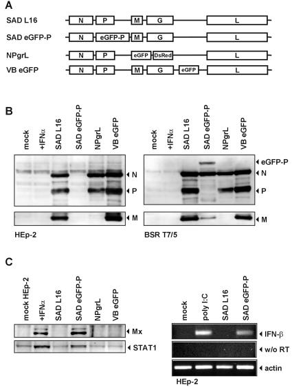
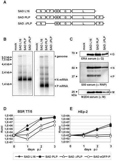
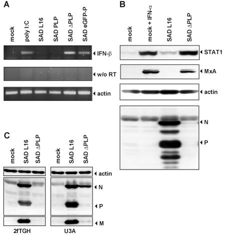
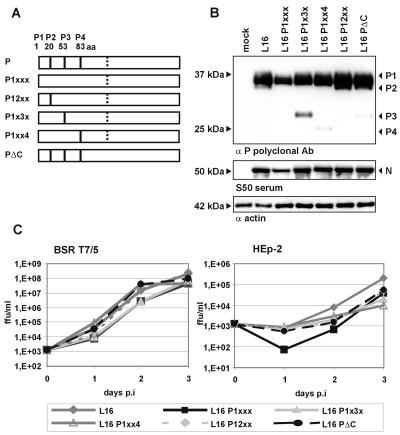
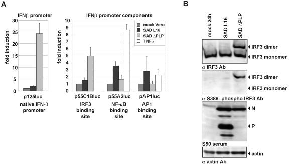
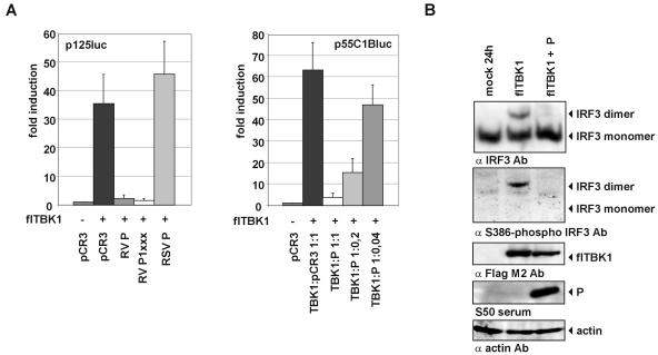
Rabies virus (RV) of the Rhabdoviridae family grows in alpha/beta interferon (IFN)-competent cells, suggesting the existence of viral mechanisms preventing IFN gene expression. We here identify the viral phosphoprotein P as the responsible IFN antagonist. The critical involvement of P was first suggested by the observation that an RV expressing an enhanced green fluorescent protein (eGFP)-P fusion protein (SAD eGFP-P) (S. Finke, K. Brzozka, and K. K. Conzelmann, J. Virol. 78:12333-12343, 2004) was eliminated in IFN-competent HEp-2 cell cultures, in contrast to wild-type (wt) RV or an RV replicon lacking the genes for matrix protein and glycoprotein. SAD eGFP-P induced transcription of the IFN-beta gene and expression of the IFN-responsive MxA and STAT-1 genes. Similarly, an RV expressing low levels of P, which was generated by moving the P gene to a promoter-distal gene position (SAD DeltaPLP), lost the ability to prevent IFN induction. The analysis of RV mutants lacking expression of truncated P proteins P2, P3, or P4, which are expressed from internal AUG codons of the wt RV P open reading frame, further showed that full-length P is competent in suppressing IFN-beta gene expression. In contrast to wt RV, the IFN-inducing SAD DeltaPLP caused S386 phosphorylation, dimerization, and transcriptional activity of IFN regulatory factor 3 (IRF-3). Phosphorylation of IRF-3 by TANK-binding kinase-1 expressed from transfected plasmids was abolished in wt RV-infected cells or by cotransfection of P-encoding plasmids. Thus, RV P is necessary and sufficient to prevent a critical IFN response in virus-infected cells by targeting activation of IRF-3 by an upstream kinase.


Figures

FIG. 1.
FIG. 1.
SAD eGFP-P induces IFN-β gene expression. (A) Genome organization of wt SAD L16; SAD eGFP-P, expressing a P protein with an N-terminal eGFP-moiety; NPgrL virus, with M and G genes replaced with eGFP and DsRed genes; and SAD VB eGFP, containing an extra eGFP gene. (B) Whole-cell extracts of BSR T7/5 and HEp-2 cells were harvested at 48 h p.i. (MOI of 1) and analyzed by Western blotting for expression of the viral N, P, and M proteins. In contrast to infection of BSR cells (right panel), SAD eGFP-P produced low levels of N and eGFP-P protein in HEp-2 cells (left panel). (C) SAD eGFP-P infection of HEp-2 cells induces transcription of IFN-β mRNA as shown by RT-PCR (right panel). and up-regulation of MxA and STAT1 proteins as shown by Western blotting (left panel).
FIG. 2.
FIG. 2.
The level of P protein is crucial for growth of RV in IFN-competent cells. (A) Genome organization of recombinant RV. SAD PLP carries an extra copy of the P gene downstream of L (gene order, 3′-N-P-M-G-L-P-5′). In SAD ΔPLP, the P gene downstream of N in SAD PLP was deleted, resulting in a virus with a single P gene copy downstream of L (gene order, 3′-N-M-G-L-P-5′). (B) Northern blot hybridization with N and P gene-specific cDNA of RNA from BSR T7/5 cells infected for 24 h at an MOI of 1 with the indicated viruses. SAD ΔPLP virus produces lower levels of P mRNA than SAD L16. (C) Expression of RV N, P, M, and G proteins was analyzed in Western blots of cell extracts from BSR T7/5 at 24 h p.i. at an MOI of 1. (D and E) Single-step growth curves were performed on BSR T7/5 cells (D) and on HEp-2 cells (E) after infection with the indicated viruses at an MOI of 0.01. ffu, focus-forming units.
FIG. 3.
FIG. 3.
The level of P protein is crucial for IFN-β gene expression. (A) SAD ΔPLP effectively induces transcription of IFN-β mRNA in HEp-2 cells as shown by RT-PCR. Mock-infected cells were stimulated by poly(I · C) transfection. RNA was isolated 20 h after infection at an MOI of 1. For RT-PCR, primers specific for IFN-β or β-actin (as a loading control) were used. (B) SAD ΔPLP infection induces an antiviral response and up-regulates expression of MxA and STAT1 as shown by Western blotting at 48 h p.i. Only in wt SAD L16-infected cells were abundant amounts of RV N and P proteins detected.(C) Infection (MOI of 1) with SAD ΔPLP of U3A cells lacking STAT-1 leads to accumulation of viral proteins, in contrast to the case for the parental STAT-1-containing 2fTGH cells.
FIG. 4.
FIG. 4.
Mutation of internal AUG start codons does not abrogate IFN-antagonistic activity of P. (A) Schematic representation of mutations introduced into the P protein. Solid vertical bars represent authentic methionine codons. Lack of the bar indicates substitution with isoleucine codons. Dotted vertical bars represent the AUG codon of the hypothetical C of P ORF + 1. In SAD PΔC, the AUG codon was mutated to ACG. (B) Protein synthesis of RV encoding mutated P proteins. BSR T7/5 cells were infected with the indicated viruses at an MOI of 1. Cell extracts were analyzed by Western blotting at 24 h p.i. P and N proteins were detected by mouse polyclonal anti-P serum and rabbit polyclonal serum S50, respectively. (C) Single-step growth curves of RVs lacking short forms of P protein on BSR T7/5 and HEp-2 cells infected at an MOI of 0.01. RV SAD P1xxx, lacking P2, P3, and P4, grew productively in HEp-2 cells.
FIG. 5.
FIG. 5.
Rabies virus targets the IRF-3 activation pathway. (A) Vero cells were transfected with reporter plasmid p125luc, p55C1Bluc, p55A2luc, or pAP1luc and infected at an MOI of 3 with SAD L16 or SAD ΔPLP. Cell lysates were analyzed using the dual luciferase reporter system (Promega) for luciferase activity at 48 h p.i. In contrast to SAD ΔPLP, SAD L16 is able to prevent expression of firefly luciferase from plasmids containing the IFN-β promoter (p125luc) and the IRF-3 binding site (p55C1Bluc). In contrast to IRF-3, the activities of NF-κB and AP-1 are not further stimulated in SAD ΔPLP-infected cells compared to SAD L16-infected cells. Incubation of cells with tumor necrosis factor alpha was used as a positive control. Error bars indicate standard deviations. (B) Dimerization and Ser386 phosphorylation of IRF-3 in SAD ΔPLP-infected cells. HEp-2 cells were infected at an MOI of 1 and cell extracts were analyzed at 24 h p.i. by native PAGE and Western blotting. In contrast to SAD ΔPLP, wt SAD L16 prevents IRF-3 dimerization and phosphorylation on Ser386. The same cell lysates were analyzed by SDS-PAGE for expression of viral N and P proteins.
FIG. 6.
FIG. 6.
Expression of P is sufficient to prevent TBK-1-mediated IFN-β induction. (A) HEK 293 cells were transfected with expression plasmids encoding the indicated genes and reporter plasmids harboring the firefly luciferase gene under control of the IFN-β promoter and renilla luciferase controlled by the CMV promoter. Luciferase activities were determined at 48 h posttransfection. Cotransfection of P or of P1xxx with TBK-1 inhibited activation of the IFN-β promoter almost completely. The P protein of BRSV (RSV P) was used as a control that is not able to inhibit TBK-1-mediated activation of the p125luc reporter plasmid (left panel). The inhibition of TBK-1-mediated activation of IRF-3 by RV P is dose dependent as revealed by luciferase expression from p55C1Bluc (right panel). Error bars indicate standard deviations. (B) Expression of RV P inhibits TBK-1-mediated dimerization of endogenous IRF-3 and phosphorylation of IRF-3 at Ser386. Cell extracts were harvested at 24 h posttransfection of TBK-1 or TBK-1 and P-encoding plasmids and were analyzed by native PAGE and Western blotting. Expression of Flag-tagged TBK-1 (fl-TBK1), RV P, and β-actin was confirmed by SDS-PAGE and Western blot analysis of the same lysates.

Similar articles

Cited by

References

    1. Ahmed, M., M. O. McKenzie, S. Puckett, M. Hojnacki, L. Poliquin, and D. S. Lyles. 2003. Ability of the matrix protein of vesicular stomatitis virus to suppress beta interferon gene expression is genetically correlated with the inhibition of host RNA and protein synthesis. J. Virol. 77:4646-4657. - PMC - PubMed
    1. Akira, S., and K. Takeda. 2004. Toll-like receptor signalling. Nat. Rev. Immunol. 4:499-511. - PubMed
    1. Basler, C. F., and A. Garcia-Sastre. 2002. Viruses and the type I interferon antiviral system: induction and evasion. Int. Rev. Immunol. 21:305-337. - PubMed
    1. Basler, C. F., A. Mikulasova, L. Martinez-Sobrido, J. Paragas, E. Muhlberger, M. Bray, H. D. Klenk, P. Palese, and A. Garcia-Sastre. 2003. The Ebola virus VP35 protein inhibits activation of interferon regulatory factor 3. J. Virol. 77:7945-7956. - PMC - PubMed
    1. Basler, C. F., X. Wang, E. Muhlberger, V. Volchkov, J. Paragas, H. D. Klenk, A. Garcia-Sastre, and P. Palese. 2000. The Ebola virus VP35 protein functions as a type I IFN antagonist. Proc. Natl. Acad. Sci. USA 97:12289-12294. - PMC - PubMed

Publication types

MeSH terms

LinkOut - more resources