. 2005 Jun;79(12):7682-97.
doi: 10.1128/JVI.79.12.7682-7697.2005.

Single amino acid insertions at the junction of the sindbis virus E2 transmembrane domain and endodomain disrupt virus envelopment and alter infectivity

Affiliations

Single amino acid insertions at the junction of the sindbis virus E2 transmembrane domain and endodomain disrupt virus envelopment and alter infectivity

Raquel Hernandez et al. J Virol. 2005 Jun.

Abstract

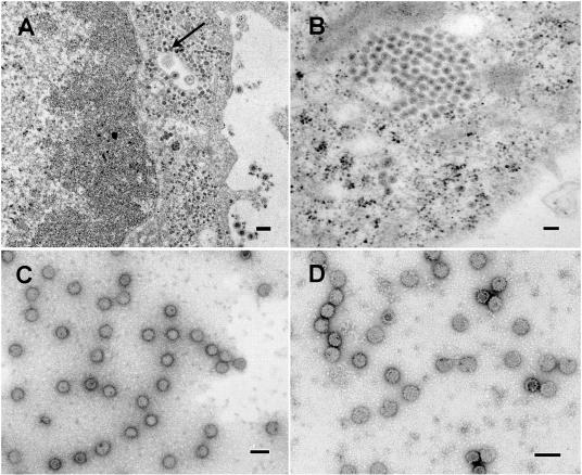
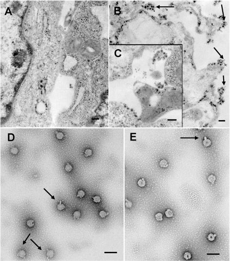
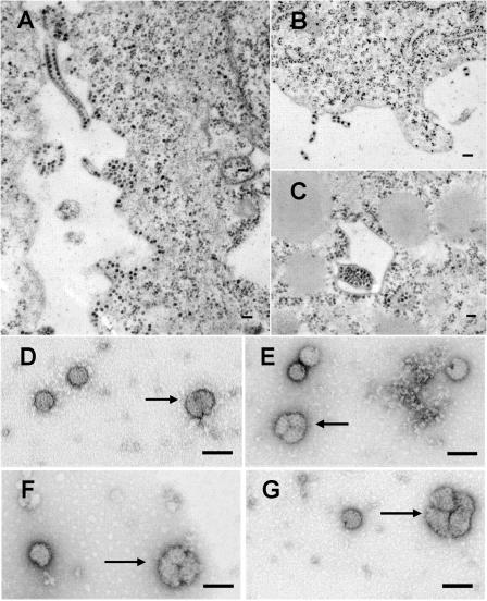
The final steps in the envelopment of Sindbis virus involve specific interactions of the E2 endodomain with the virus nucleocapsid. Deleting E2 K at position 391 (E2 DeltaK391) resulted in the disruption of virus assembly in mammalian cells but not insect cells (host range mutant). This suggested unique interactions of the E2 DeltaK391 endodomain with the different biochemical environments of the mammalian and insect cell lipid bilayers. To further investigate the role of the amino acid residues located at or around position E2 391 and constraints on the length of the endodomain on virus assembly, amino acid insertions/substitutions at the transmembrane/endodomain junction were constructed. An additional K was inserted at amino acid position 392 (KK391/392), a K-->F substitution at position 391 was constructed (F391), and an additional F was inserted at 392 (FF391/392). These changes should lengthen the endodomain in the KK391/392 insertion mutant or shorten the endodomain in the FF391/392 mutant. The mutant FF391/392 grown in BHK cells formed virus particles containing extruded material not found on wild-type virus. This characteristic was not seen in FF391/392 virus grown in insect cells. The mutant KK391/392 grown in BHK cells was defective in the final membrane fission reaction, producing multicored or conjoined virus particles. The production of these aberrant particles was ameliorated when the KK391/392 mutant was grown in insect cells. These data indicate that there is a critical minimal spanning distance from the E2 membrane proximal amino acid at position 391 and the conserved E2 Y400 residue. The observed phenotypes of these mutants also invoke an important role of the specific host membrane lipid composition on virus architecture and infectivity.


Figures

FIG. 1.
FIG. 1.
Electron micrographs of Sindbis virus-infected BHK and insect U4.4 cells. Panels A and B show thin sections of wild-type infections in BHK cells (A) and in insect U4.4 cells (B). In panel A, the arrow highlights a region within the cell containing many nucleocapsids associated with internal membranes. Virus particles are also seen budding from the plasma membrane. In panel B, mature virus particles are seen within internal vesicles typical of wild-type virus assembly in insect cells. Panels C and D are negative stains of gradient-purified wild-type virus particles grown in BHK cells (C) or insect cells (D). Virus particles from both host cells are symmetrical, displaying few empty particles. Bars, 100 nm.
FIG. 2.
FIG. 2.
Electron micrographs of BHK cells infected with the mutant FF391/392. In panel A, nucleocapsids are seen distributed through the cell cytoplasm as well as associated with the membrane. The arrows in panel B indicate the types of budding virus particles seen at the cell plasma membrane. These virus particles appear associated with extraneous nonvirus material, which are found clumped together at the plasma membrane, also shown in panel C. Bars for panels A to C, 200 nm. Negatively stained preparations of gradient-purified mutant virus shown in panels D and E illustrate the abnormal structure of particles produced from this mutant. Of note (arrows) are the “lollypop” and “mouse ear” appendages seen on most of the particles in addition to the more amorphous appearance of these particles. Bars for panels D and E, 100 nm.
FIG. 3.
FIG. 3.
Electron micrographs of BHK cells infected with the mutant KK391/392. As seen in panel A, this mutant produces wild-type levels of nucleocapsids seen within the cytoplasm and found associated with cell membranes. This mutant, however, produces long tubes of membrane containing multiple associated cores. Seen in panel B are virus particles containing 2, 3, and 6 cores budding from the cell surface. In panel C, a large bolus of virus cores is seen budding within an internal vesicle, surrounded by membranes associated with virus cores. In panels D to G, negatively stained purified mutant virus is seen forming conjoined particles containing 2 (D), 3 (E, F), and 4 lobes (G). Bars, 100 nm.
FIG. 4.
FIG. 4.
Electron micrographs of BHK-grown F391 mutant. Panels A and B are representative of infected BHK cells. In panel B, many particles are seen budding directly from the cell membrane. In panels C and D, negatively stained gradient-purified virus from BHK cells display normal spherical virus particles with a few empty particles visible. Bars, 100 nm.
FIG. 5.
FIG. 5.
Electron micrographs of FF391/392 mutant-infected insect U4.4 cells. In panel A, large internal vesicles containing matured virus are visible, while in panel B, particles are seen budding from the plasma membrane. Both panels A and B show particles with a wild-type appearance. Panels C to G display negative stains of gradient-purified mutant virus. Arrows in panels D to F highlight a few larger particles that are formed by this mutant in the insect cell host. Compared with mutant virus particles produced from BHK cells (Fig. 2D and E), the virus morphology of the FF391/392 mutant grown in U4.4 cells is more similar to the wild-type virus (Fig. 1D). Bars, 100 nm.
FIG. 6.
FIG. 6.
Electron micrographs of insect U4.4 cells infected with the mutant KK391/392. In panel A, a cytoplasmic vesicle containing matured virus is shown. In panel B, the inset shows budding virus particles with visible membrane stalks (arrow), indicative of an aberrant membrane fission process. Panel C demonstrates long tubes of multicored membrane appendages also seen produced by this virus grown in BHK cells (Fig. 3A, B, and C). Negative stains of membrane-purified virus reveal a different phenotype than that seen for this mutant grown in BHK cells (Fig. 3D to G). (D to F) Virus grown in U4.4 cells appears pocked and fragile (arrows). Bars, 100 nm.
FIG. 7.
FIG. 7.
Electron micrographs of insect U4.4 cells infected with the mutant F391. In panels A and B, mature virus particles are seen. In the negative stains shown in panels C and D, gradient-purified virus particles are normal in appearance (compare to Fig. 1D). Bars, 100 nm.
FIG. 8.
FIG. 8.
Isopycnic sedimentation and analysis of wild-type virus and endodomain mutant virus particles grown in BHK cells. Panel A is a graph of the sedimentation profile of mutants and wild-type virus through potassium tartrate as detailed in Materials and Methods. Wild-type virus (♦, black arrow) produced a peak of virus in fraction 3, displaying a small shoulder at fraction 4 (black arrow). The F391 mutant (▪) also gave a virus peak at fraction 3, similar to that of wild-type virus, without the virus shoulder at fraction 4. The FF391/392 virus peak (▴, gray arrow) is seen in fraction 4 cosedimenting with the wild-type virus shoulder. The mutant virus KK391/392 (•, gray arrow) displayed the lowest density, forming a peak in fraction 5. In panel B are shown the viruses from panel A correlating the peak of labeled virus from the gradient with virus infectivity. Peak infectivity for wild-type virus (♦) corresponds to the radiolabeled peak of virus (2.3 × 10¹⁰ PFU/ml) in fraction 3, with the shoulder at fraction 4 (1.0 × 10⁷ PFU/ml). The peak virus fraction for the mutant F391 (▪), 1.3 × 10⁹ PFU/ml, corresponded with that of the wild-type virus infectivity peak in fraction 3. The FF391/392 mutant also gave a peak infectivity titer (7.0 × 10⁷ PFU/ml) in the same fraction, fraction 4, as the radioactively labeled Y420 shoulder shown in panel A. The KK391/392 mutant displayed a broad peak of infectivity beginning with fraction 4 (3.7 × 10⁴ PFU/ml), with peak infectivity seen in fraction 5 (1.0 × 10⁶ PFU/ml) compared to the peak of labeled (A) Y420. Purified viruses from panel A were used for the negative stains seen in all figures.
FIG. 9.
FIG. 9.
Fusion properties of mutant virus glycoproteins. In panel A are shown the FFWI profiles of the endodomain mutants compared to that of the wild type. Wild-type virus (⋄), the KK391/392 mutant (○), and the F391 mutant (▪) displayed the most similar profiles. The most interesting profile was seen with the FF391/392 mutant (▴), which did not achieve 100% fusion until pH 8.0. Mock-infected cells (+) displayed negligible fusion. Shown in panel B, FFWO was utilized to measure the fusion profiles of wild-type and mutant viruses grown in insect U4.4 cells with BHK cells as described in Materials and Methods. Unlike the FFWI results using BHK infected cells, insect-grown virus of all the endodomain mutants gave subtle differences in their response to the pH treatments The mutant KK391/392 (○) produced a fusion profile most similar to the FF391/392 mutant (▴), differing by a slight shift in fusion capacity. In general, viruses grown from insect U4.4 cells all appeared wild type in the pH region from pH 7.2 to pH 7.6. As in panel A, monolayers without added virus (+), treated as the virus-containing samples described above, showed negligible fusion.

Similar articles

Cited by

References

    1. Anthony, R. P., and D. T. Brown. 1991. Protein-protein interactions in an alphavirus membrane. J. Virol. 65:1187-1194. - PMC - PubMed
    1. Anthony, R. P., A. M. Paredes, and D. T. Brown. 1992. Disulfide bonds are essential for the stability of the Sindbis virus envelope. Virology 190:330-336. - PubMed
    1. Braun, P., and G. von Heijne. 1999. The aromatic residues Trp and Phe have different effects on the positioning of a transmembrane helix in the microsomal membrane. Biochemistry 38:9778-9782. - PubMed
    1. Bretscher, M. S. 1993. Cholesterol and the Golgi apparatus. Science 261:1280. - PubMed
    1. Brown, D. T., M. R. F. Waite, and E. R. Pfefferkorn. 1972. Morphology and morphogenesis of Sindbis virus as seen with freeze-etching techniques. J. Virol. 10:534-536. - PMC - PubMed

Publication types

Substances

LinkOut - more resources