. 2005 Jun;79(12):7745-55.
doi: 10.1128/JVI.79.12.7745-7755.2005.

The structure of the poliovirus 135S cell entry intermediate at 10-angstrom resolution reveals the location of an externalized polypeptide that binds to membranes

Affiliations

The structure of the poliovirus 135S cell entry intermediate at 10-angstrom resolution reveals the location of an externalized polypeptide that binds to membranes

Doryen Bubeck et al. J Virol. 2005 Jun.

Abstract

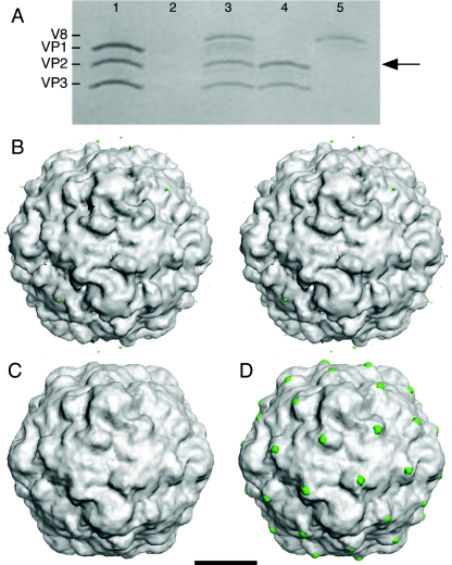
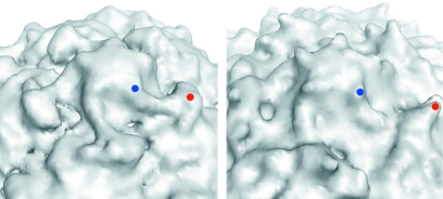
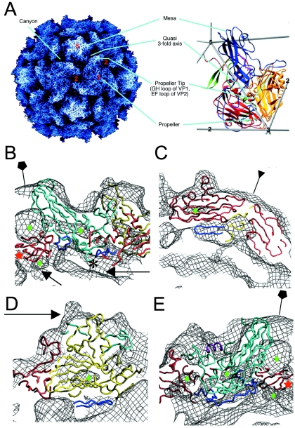
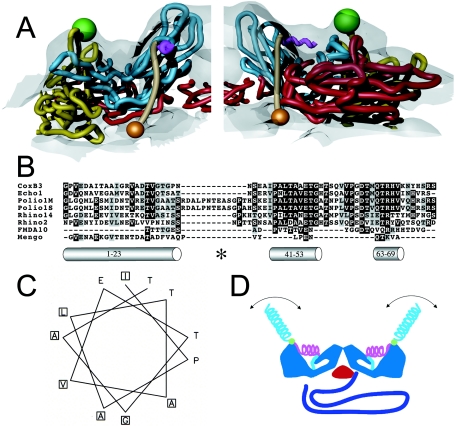
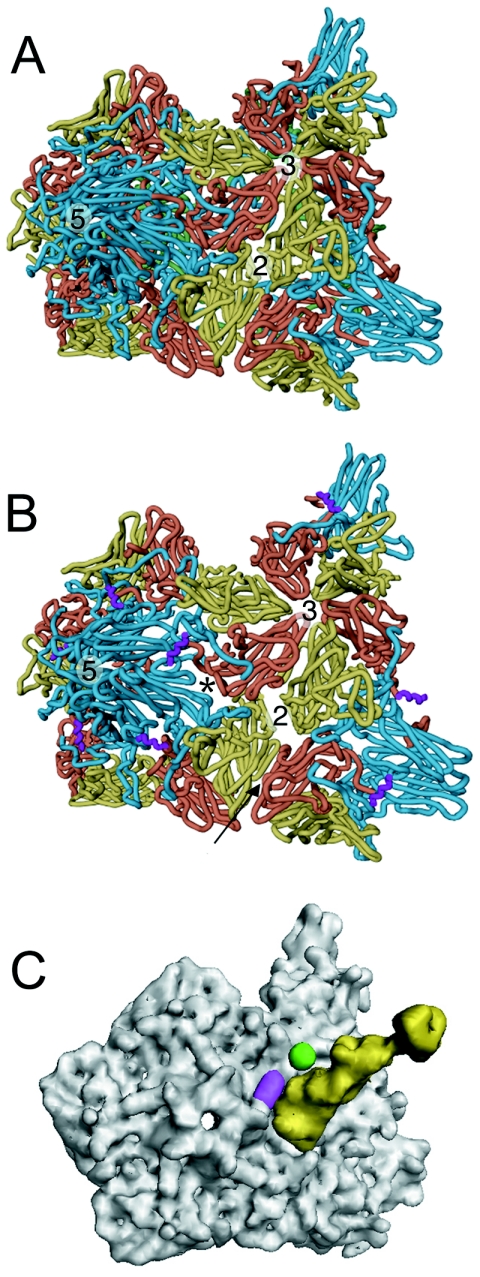
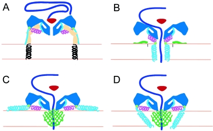
Poliovirus provides a well-characterized system for understanding how nonenveloped viruses enter and infect cells. Upon binding its receptor, poliovirus undergoes an irreversible conformational change to the 135S cell entry intermediate. This transition involves shifts of the capsid protein beta barrels, accompanied by the externalization of VP4 and the N terminus of VP1. Both polypeptides associate with membranes and are postulated to facilitate entry by forming a translocation pore for the viral RNA. We have calculated cryo-electron microscopic reconstructions of 135S particles that permit accurate placement of the beta barrels, loops, and terminal extensions of the capsid proteins. The reconstructions and resulting models indicate that each N terminus of VP1 exits the capsid though an opening in the interface between VP1 and VP3 at the base of the canyon that surrounds the fivefold axis. Comparison with reconstructions of 135S particles in which the first 31 residues of VP1 were proteolytically removed revealed that the externalized N terminus is located near the tips of propeller-like features surrounding the threefold axes rather than at the fivefold axes, as had been proposed in previous models. These observations have forced a reexamination of current models for the role of the 135S particle in transmembrane pore formation and suggest testable alternatives.


Figures

FIG. 1.
FIG. 1.
V8 protease cleaves the exposed N terminus of VP1 at the propeller tip of 135S. (A) Coomassie blue-stained sodium dodecyl sulfate-polyacrylamide gel whose lanes represent different stages in the preparation of V8-cleaved 135S. (Lane 1) The 135S particle before digestion showing the migration of the capsid proteins VP1, VP2, and VP3. (Lane 2) Blank. (Lane 3) The 135S particle after digestion with the V8 protease. Cleaved VP1 comigrates with VP2 (arrow) (14). (Lane 4) An aliquot from the void volume from a Sephacryl S-100 column demonstrating the homogeneity of the purified V8-135S particles. (Lane 5) An aliquot from the inclusion volume of the S-100 column (50 ml after the void volume peak) containing the V8 protease. (B to D) Surface renderings of poliovirus cell entry intermediate 135S and difference images (scale bar, 100 Å). Difference electron density maps between reconstructions were contoured at 2.5 σ, while both 135S and V8-cleaved 135S reconstructions were contoured at 0.75 σ (σ is the root-mean-square deviation of the electron density values). Figures were produced with DINO (http://www.dino3d.org). (B) A stereo representation of the 135S1 cryo-EM reconstruction (grey). Differences between the 135S2 and 135S1 reconstructions (green) were insignificant. The ridge between the mesa and propeller tip is labeled (arrowhead). (C) A reconstruction of the V8-proteolyzed 135S (grey) particle. A difference electron density map calculated between two V8-cleaved 135S reconstructions showed no visible differences on the outer surface. (D) Significant difference density features were seen when each V8-cleaved 135S map (grey) was subtracted from either unmodified 135S particle reconstruction. The largest differences (green) were located at the propeller tip.
FIG. 2.
FIG. 2.
Close-up view of the surface of the 135S reconstruction (left) and of native poliovirus (right) in the vicinity of the fivefold axis. Electron density for the native virus was calculated to 11-Å resolution from the atomic coordinates. In each panel, one propeller tip (red dot) and a nearby point of the fivefold mesa (blue dot) are indicated. In the 135S reconstruction a prominent linear ridge connects the mesa with the propeller and rises to a substantial elevation above the floor of the canyon. No such feature is observed in the native virus.
FIG. 3.
FIG. 3.
Pseudoatomic modeling of the 135S particle. (A) The structure of the 160S particle serves as a reference for prominent structural features. (Left panel) A radially depth-cued rendering of the atomic model (22) of the 160S particle. (Right panel) An expanded representation of a single protomer showing ribbon diagrams of VP1 (blue), VP2 (yellow), VP3 (red), and VP4 (green) overlaid on an icosahedral framework. The fivefold, threefold, and twofold axes are indicated by numbers. Cyan lines point to prominent surface features in the two panels. (B to E) Pseudoatomic models of VP1 (cyan), VP2 (yellow), and VP3 (red) resulting from rigid-body docking and refinement of native virus coordinates into the 135S density maps (black mesh). Residues outside the β-barrel cores that were not modeled previously (3) were included in the refinement (dark blue). Green diamonds signify low-density regions within the core of protein subunits. (B) A model-density overlay in the vicinity of a fivefold axis (pentagon). The RNA and protein shells make contacts (arrows) at the fivefold axis near the VP3 β tube (red star) and at the base of the canyon. The truncated N terminus of VP1 (residue 71) is indicated (*). (C) A model-density overlay in the vicinity of a threefold axis (inverted triangle). Loops at the narrow ends of the VP3 β barrel and the β barrels from symmetry-related VP2s and VP3s alternate around the threefold axis. The newly modeled VP2 β hairpin (dark blue) is seen to contact VP3. (D) Model-density overlay in the vicinity of the propeller tip (arrow) showing VP2, the GH loop and C terminus of VP1 (cyan), and the newly modeled VP2 β hairpin (dark blue) from panel C fitting into a crevice in the EM envelope. (E) A slice through the model-density overlay of a protomer. A helix occupying the external linear ridge, possibly from the residue range 41 to 53 of VP1, is shown in magenta. The newly modeled VP3 N terminus (residues 14 to 49, dark blue) can be seen within the density envelope on the underside of the capsid in panels B and E.
FIG. 4.
FIG. 4.
Localization and characterization of the N terminus of VP1. (A) Two views of 135S model coordinates, VP1 (cyan), VP2 (yellow), and VP3 (red), are overlaid on the cryo-EM 135S reconstruction (grey semitransparent). Trajectory for the N-terminal 70 residues of VP1 (tan) begins at residue 71 (orange sphere), emerges from a gap between fivefold symmetry-related copies of VP1, forms a helix (magenta) through the ridge, and connects to the V8-cleavage site (green sphere). The helix originates at the points of the star-shaped mesa. For clarity, only one protomer is shown in each panel. (B) Sequence alignment of the N-terminal extension of VP1 from different picornaviruses (coxsackievirus B3, echovirus 1, poliovirus 1 Mahoney strain, poliovirus 1 Sabin strain, rhinovirus 14, rhinovirus 2, foot-and-mouth-disease virus 10, and mengovirus, top to bottom, respectively) reveals a highly conserved stretch of residues (41 to 53, mostly dark highlighting) that is predicted by the Chou-Fasman algorithm to include an alpha helix. The algorithm also identified the extreme N-terminal 20 residues as a helix. This amphipathic helix was previously identified, and it was proposed to form a pore in the host cell membrane (14). A third helical stretch, located near residue 70, is observed in the native virus making contacts with the VP1 β barrel. All predicted helices are depicted as grey cylinders and labeled with their respective residue numbers. The V8 cleavage site is labeled (*). (C) A helical wheel representation (generated using EMBOSS Pepwheel, www.hgmp.mrc.ac.uk/Software/EMBOSS/Apps/pepwheel.html) of VP1 residues 41 to 52 reveals an amphipathic arrangement of side chains. (D) Schematic of the externalization of the VP1 N terminus showing the capsid (blue), VP3 β tube (red), RNA (purple), and the N terminus of VP1 (residues 71 to 54 [cyan], 53 to 41 [magenta helix], V8 cleavage site [green sphere], and 31 to 1 [cyan helix]). The N-terminal 20 residues that are thought to form an amphipathic helix (14) could be either disordered or mobile in the absence of lipid (cyan helix).
FIG. 5.
FIG. 5.
Comparison of 135S with native virus. The transition from native poliovirus (A) to the 135S intermediate (B) is characterized by an expansion of the virus capsid causing gaps in the model between symmetry-related copies of the protomer. The most pronounced openings occur between VP1 and VP3 at the base of the canyon (*) and at the twofold axis. VP1, VP2, and VP3 are cyan, yellow, and red, respectively. In the 135S model, the helix proposed to occupy the external linear ridge is magenta; in native virus, VP4 is green. The arrow indicates the six-stranded β sheet formed by the hairpin of VP2 and the four-stranded β sheet from VP3 of a different, symmetry-related pentamer. (C) Electron density calculated for Pvr (yellow) overlaid with density calculated for the pseudoatomic 135S model (grey) does not clash with the ridge corresponding to the externalized N terminus of VP1 (magenta) or with the V8 cleavage site (green).
FIG. 6.
FIG. 6.
Working models for poliovirus entry. A cross section of a portion of the capsid is shown in dark blue, VP4 is green, and the N terminus of VP1 is cyan and magenta. The V8 cleavage site is shown as a green circle. (A) Native poliovirus binds its receptor, Pvr (ectodomains 1 to 3, tan; transmembrane domain, black helix), and at physiological temperature undergoes an irreversible change to the 135S particle. The path of egress of the N terminus of VP1, suggested by the present 135S reconstruction, would not preclude the continued binding of Pvr. At this stage, the VP3 β tube (red) blocks an otherwise open channel along the fivefold axis. (B to D) Alternative models for the direct anchoring of the virus to the membrane via the N terminus of VP1 and formation of a transmembrane pore for RNA translocation. To accommodate the passage of RNA (purple), the VP3 β tube has shifted, and the channel has expanded, becoming continuous with a pore through the membrane. The V8 cleavage site remains accessible. (B) Amphipathic helices at the N terminus of VP1 (cyan) may form a five-helix bundle close to the fivefold axis, which would require the magenta helix to dissociate from the body of the virus. Alternatively, VP4 may play a more central role in pore formation (C and D). In that case, VP1 may participate directly in forming the pore (D) or serve as a nonspecific membrane anchor (C).

References

    1. Baker, T. S., and R. H. Cheng. 1996. A model-based approach for determining orientations of biological macromolecules imaged by cryoelectron microscopy. J. Struct. Biol. 116:120-130. - PubMed
    1. Basavappa, R., R. Syed, O. Flore, J. P. Icenogle, D. J. Filman, and J. M. Hogle. 1994. Role and mechanism of the maturation cleavage of VP0 in poliovirus assembly: structure of the empty capsid assembly intermediate at 2.9Å resolution. Protein Sci. 3:1651-1669. - PMC - PubMed
    1. Belnap, D. M., D. J. Filman, B. L. Trus, N. Cheng, F. P. Booy, J. F. Conway, S. Curry, C. N. Hiremath, S. K. Tsang, A. C. Steven, and J. M. Hogle. 2000. Molecular tectonic model of virus structural transitions: the putative cell entry states of poliovirus. J. Virol. 74:1342-1354. - PMC - PubMed
    1. Belnap, D. M., W. D. Grochulski, N. H. Olson, and T. S. Baker. 1993. Use of radial density plots to calibrate image magnification for frozen-hydrated specimens. Ultramicroscopy 48:347-358. - PMC - PubMed
    1. Belnap, D. M., B. M. McDermott, Jr., D. J. Filman, N. Cheng, B. L. Trus, H. J. Zuccola, V. R. Racaniello, J. M. Hogle, and A. C. Steven. 2000. Three-dimensional structure of poliovirus receptor bound to poliovirus. Proc. Natl. Acad. Sci. USA 97:73-78. - PMC - PubMed

Publication types