. 2005 Jun;79(12):7803-11.
doi: 10.1128/JVI.79.12.7803-7811.2005.

Allosteric effects of ligands and mutations on poliovirus RNA-dependent RNA polymerase

Affiliations

Allosteric effects of ligands and mutations on poliovirus RNA-dependent RNA polymerase

Joanna E Boerner et al. J Virol. 2005 Jun.

Abstract

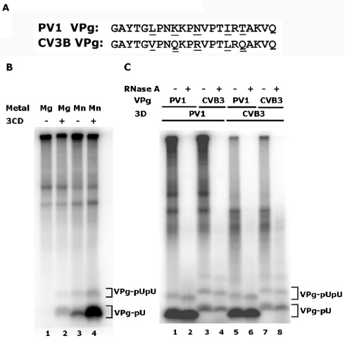
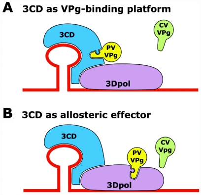
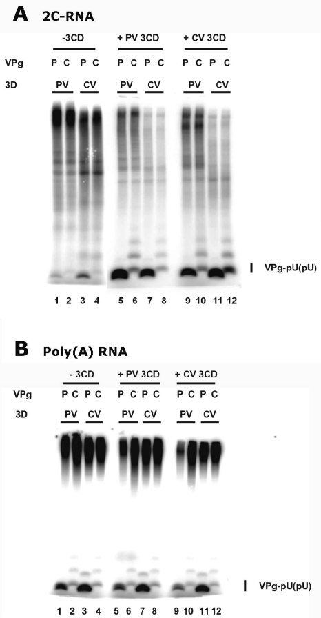
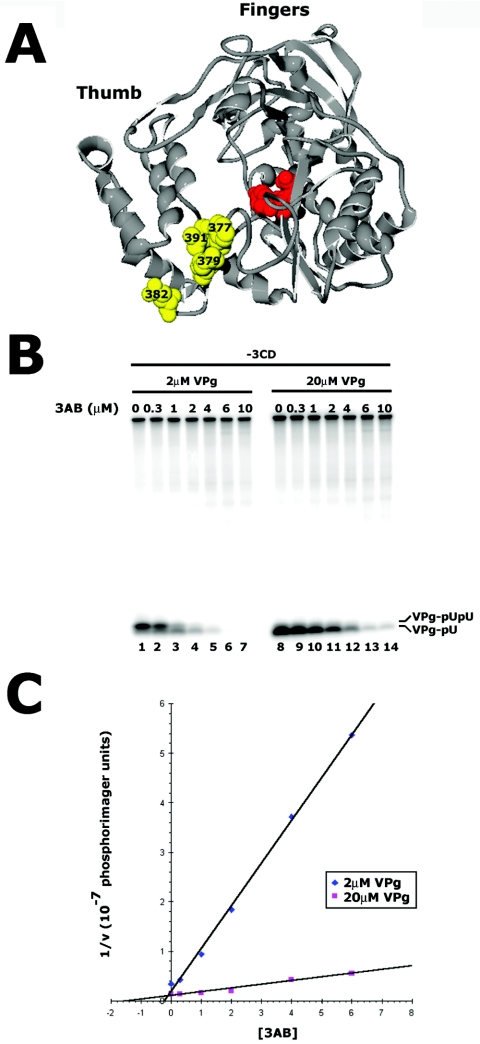
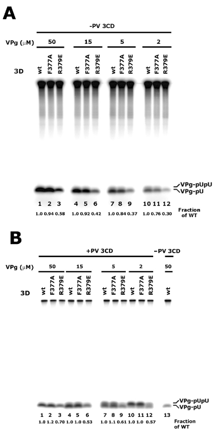
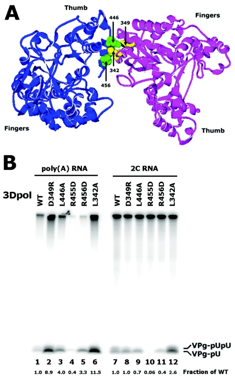
Protein priming of viral RNA synthesis plays an essential role in the replication of picornavirus RNA. Both poliovirus and coxsackievirus encode a small polypeptide, VPg, which serves as a primer for addition of the first nucleotide during synthesis of both positive and negative strands. This study examined the effects on the VPg uridylylation reaction of the RNA template sequence, the origin of VPg (coxsackievirus or poliovirus), the origin of 3D polymerase (coxsackievirus or poliovirus), the presence and origin of interacting protein 3CD, and the introduction of mutations at specific regions in the poliovirus 3D polymerase. Substantial effects associated with VPg origin were traced to differences in VPg-polymerase interactions. The effects of 3CD proteins and mutations at polymerase-polymerase intermolecular Interface I were most consistent with allosteric effects on the catalytic 3D polymerase molecule. In conclusion, the efficiency and specificity of VPg uridylylation by picornavirus polymerases is greatly influenced by allosteric effects of ligand binding that are likely to be relevant during the viral replicative cycle.


Figures

FIG. 1.
FIG. 1.
VPg uridylylation catalyzed by poliovirus (PV-1) or coxsackievirus B3 (CVB3) 3D polymerase using a poliovirus 2C RNA template in the presence of poliovirus 3CD. (A) The sequences of PV-1 and CVB3 VPg proteins are shown. Differences in sequence are underlined. (B) PV-1 VPg uridylylation by PV-1 3D polymerase using a 2C RNA template in the presence of magnesium acetate (lanes 1 and 2) or manganese chloride (lanes 3 and 4). Poliovirus 3CD was absent in lanes 1 and 3 and present in lanes 2 and 4. (C) VPg uridylylation by PV 3D polymerase (lanes 1 to 4) and CVB3 3D polymerase (lanes 5 to 8) using a 2C RNA template; all reactions contained PV 3CD. The identity of the VPg substrate, from PV or CVB3, is indicated. Products were digested with RNase A (100 μg/ml, 30°C, 60 min) in even-numbered lanes as indicated. Products were resolved in 12% polyacrylamide-Tris-Tricine gels. The positions of migration of VPg-pUpU and VPg-pU are indicated in both (B) and (C). The identity of VPg-pU and VPg-pUpU were confirmed by RNase and snake venom phosphodiesterase treatment; slightly greater differences in mobility are seen in this gel than in subsequent separations of VPgpU and VPgpUpU.
FIG. 2.
FIG. 2.
Models for the mechanism by which 3CD stimulates VPg uridylylation by 3D polymerase. (A) In the “3CD as VPg binding platform” model, PV VPg preferentially binds to a similar binding site on both PV and CV 3CD. (B) In the “3CD as allosteric effector” model, 3CD binding to 3D causes a conformational change in either PV or CV 3D polymerase, modifying its uridylylation activity, VPg binding affinity, or both. During the catalysis of VPg uridylyation, the scenarios in both (A) and (B) must include additional contacts from those pictured, including the proximity of the active site of the 3D polymerase, the templating A residues in the 2C RNA, and the uridylylated tyrosine of the VPg molecule.
FIG. 3.
FIG. 3.
VPg uridylylation by PV and CVB3 3D polymerase using two different RNA templates in the presence and absence of poliovirus or coxsackievirus 3CD. The relative rates of uridylylation of poliovirus (P) or coxsackievirus (C) VPg by poliovirus (PV) or coxsackievirus (CV) polymerase are reflected by the intensity of the uridylylated VPg bands. Lanes 1 to 4 show the results of reactions performed in the absence of 3CD; results shown in lanes 5 to 8 were performed in the presence of PV 3CD, and results shown in lanes 9 to 12 were performed in the presence of CV 3CD. (A) 2C RNA template and (B) Poly(A) template. Products were resolved in a 12% polyacrylamide-Tris-Tricine gel and analyzed as in Fig. 1.
FIG. 4.
FIG. 4.
Inhibition of PV VPg uridylylation by poliovirus 3AB. (A) A representation of the “back of the palm” region of the full-length poliovirus polymerase structure (40) is shown. Residues F377, R379, E382, and V391, known to be involved in 3AB binding to 3D polymerase, are identified in yellow. The active site is shown in red. (B) The effects on VPg uridylylation of increasing concentrations of 3AB at two VPg concentrations (2 μM, lanes 1 to 7; 20 μM VPg, lanes 8 to 14) with a 2C RNA template are shown. Products were resolved in a 12% polyacrylamide-Tris-Tricine gel as previously. (C) Graphical analysis of the data from panel B. A plot of 1/V as a function of inhibitor (3AB) concentration shows a y-intercept above the x-axis, arguing that the inhibition of VPg uridylylation by 3AB is competitive (30). An apparent Ki of approximately 100 nM was determined by extrapolation of the intercept of the two lines to the x-axis. Velocity, V, is in phosphorimager units per unit time.
FIG. 5.
FIG. 5.
The effect of mutations in the 3AB binding cluster on the rate of VPg uridylylation in the absence and presence of 3CD. (A). The rates of poliovirus VPg uridylyation by the wild type, F377A and R379E poliovirus 3D polymerases were measured at four different VPg concentrations in the absence of poliovirus 3CD as indicated. The fraction of wild-type activity displayed by the mutant polymerases is shown. (B). The extent of uridylylation in the presence of PV 3CD was assessed as described for panel A; the exposure shown, however, is one-tenth that shown in (A). For both panels, products were analyzed in 12% polyacrylamide-Tris-Tricine gels, as described previously.
FIG. 6.
FIG. 6.
The effect of mutations at Interface I in 3D polymerase on VPg uridylylation. (A) Two full-length polymerase monomers (40), interacting at Interface I identified from the wild-type polymerase structure (12) are shown. The “thumb” and “fingers” regions of each polymerase are marked to provide orientation, and the residues that were mutated individually in various polymerases are labeled. Residues L446, D455, and D456 on the “thumb” of the polymerase on the left are shown in green; residues L342 and D349 on the back of the “palm” region of the polymerase on the right are shown in yellow. (B) The extent of VPg uridylylation by wild-type and various 3D polymerases with mutations in residues at Interface I was measured in the presence of either poly(A) templates (1 μM strands) or 2C RNA templates (0.15 μM strands), as indicated, in the absence of poliovirus 3CD protein. The fraction of VPg uridylylation activity for each mutant polymerase, compared to the wild type, is shown. Products were resolved in 12% polyacrylamide-Tris-Tricine gels and analyzed as described in Materials and Methods. The radioactivity often present in the wells represents, for the most part, 3′-labeled template RNA, due to the terminal transferase activity of polymerase, as evidenced by the reduction of this signal when template RNA with a blocked 3′-hydroxyl was used (data not shown). The positions of migration of VPg-pUpU and VPg-pU are indicated.

References

    1. Bell, Y. C., B. L. Semler, and E. Ehrenfeld. 1999. Requirement for RNA replication of a poliovirus replicon by coxsackievirus B3 RNA polymerase. J. Virol. 73:9413-9421. - PMC - PubMed
    1. Cheetham, G. M. T., and T. A. Steitz. 1999. Structure of a transcribing T7 RNA polymerase initiation complex. Science 286:2305-2309. - PubMed
    1. Cornell, C. T., R. Perera, J. E. Brunner, and B. L. Semler. 2004. Strand-specific RNA synthesis determinants in the RNA-dependent RNA polymerase of poliovirus. J. Virol. 78:4397-4407. - PMC - PubMed
    1. Crowder, S., and K. Kirkegaard. 2004. Complete three-dimensional structures of picornaviral RNA-dependent RNA polymerases. Structure 12:1336-1339. - PubMed
    1. Dayhuff, T. J., R. F. Gesteland, and J. F. Atkins. 1992. Electrophoresis, autoradiography and electroblotting of peptides: T4 gene 60 hopping. BioTechniques 13:500-503. - PubMed

Publication types

MeSH terms

LinkOut - more resources