. 2005 Jun;79(12):7827-37.
doi: 10.1128/JVI.79.12.7827-7837.2005.

Characterization of human cytomegalovirus glycoprotein-induced cell-cell fusion

Affiliations

Characterization of human cytomegalovirus glycoprotein-induced cell-cell fusion

Eric R Kinzler et al. J Virol. 2005 Jun.

Abstract

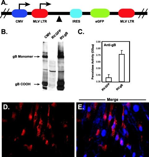
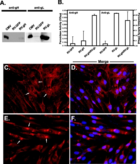
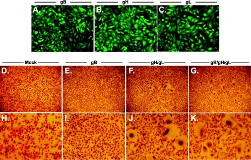
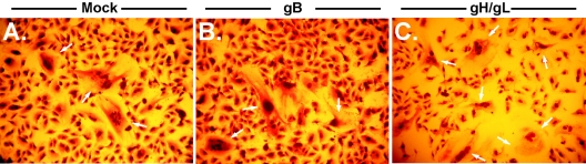
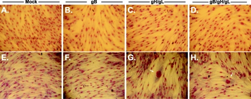
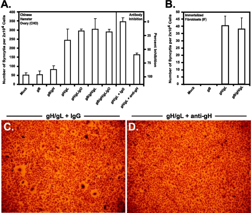
Human cytomegalovirus (CMV) infection is dependent on the functions of structural glycoproteins at multiple stages of the viral life cycle. These proteins mediate the initial attachment and fusion events that occur between the viral envelope and a host cell membrane, as well as virion-independent cell-cell spread of the infection. Here we have utilized a cell-based fusion assay to identify the fusogenic glycoproteins of CMV. To deliver the glycoprotein genes to various cell lines, we constructed recombinant retroviruses encoding gB, gH, gL, and gO. Cells expressing individual CMV glycoproteins did not form multinucleated syncytia. Conversely, cells expressing gH/gL showed pronounced syncytium formation, although expression of gH or gL alone had no effect. Anti-gH neutralizing antibodies prevented syncytium formation. Coexpression of gB and/or gO with gH/gL did not yield detectably increased numbers of syncytia. For verification, these results were recapitulated in several cell lines. Additionally, we found that fusion was cell line dependent, as nonimmortalized fibroblast strains did not fuse under any conditions. Thus, the CMV gH/gL complex has inherent fusogenic activity that can be measured in certain cell lines; however, fusion in fibroblast strains may involve a more complex mechanism involving additional viral and/or cellular factors.


Figures

FIG. 1.
FIG. 1.
Retroviral expression of gB in CHO and NHDF cells. (A) The genes encoding CMV glycoproteins were cloned into plasmid pCMMP.MCS.IRES.GFP at the multiple cloning site (▴). Transcripts are driven by either the CMV promoter or the MLV long terminal repeat (LTR) after transfection or transduction, respectively. In each case, transcripts are bicistronic, carrying both the transgene and the gene encoding GFP (eGFP) under translational control of an IRES. Recombinant retrovirus was generated as described in Materials and Methods. (B) CHO cells were transduced with a recombinant retrovirus encoding gB (RV.gB) or an empty retrovirus encoding only GFP (RV.GFP), and glycoprotein expression was confirmed by reducing 7.5% sodium dodecyl sulfate-polyacrylamide gel electrophoresis followed by immunoblotting with monoclonal antibody 27-78 (anti-gB), recognizing the carboxy-terminal region of gB. Mouse antibodies were identified with goat anti-rabbit-HRP and detected by enhanced chemiluminescence. Retrovirally transduced cells express both monomeric and cleaved (COOH) gB. In all figures, CMV represents NHDF cell lysates infected with strain AD169 at a multiplicity of infection of 1 and harvested at 6 days postinfection, RV.GFP represents CHO cells transduced with an empty retrovirus, and RV.gB represents cells transduced with a retrovirus encoding gB. (C) Cell surface expression was examined by CELISA with anti-gB antibodies on gB-expressing cells. Mouse antibodies were identified with goat anti-mouse-HRP, followed by peroxidase detection with TMB substrate. Error bars indicate standard deviations. (D and E) To visualize gB expression, NHDF cells expressing gB were stained with anti-gB antibodies and goat anti-mouse-Alexa Fluor 594. (E) In a merged photograph, blue staining represents DAPI-stained nuclei. OD450, optical density at 450 nm.
FIG. 2.
FIG. 2.
Retroviral expression of gH and gL in CHO and NHDF cells. (A) CHO cells were transduced with a retrovirus encoding gH (RV.gH) or gL (RV.gL), and expression was confirmed by reducing 10% sodium dodecyl sulfate-polyacrylamide gel electrophoresis followed by immunoblotting with polyclonal antibody 6824 (anti-gH) or 6394 (anti-gL). Rabbit antibodies were identified with goat anti-rabbit-HRP and detected by enhanced chemiluminescence. (B) Cell surface gH/gL expression was examined by CELISA with AP86 (anti-gH) or anti-gL antibodies. Mouse antibodies were detected with goat anti-mouse-HRP, while rabbit antibodies were detected with goat anti-rabbit-HRP followed by peroxidase detection with TMB substrate. Error bars indicate standard deviations. (C to F) Glycoprotein expression was visualized by immunofluorescence with AP86 (anti-gH, panels C and D) or anti-gL (panels E and F). In each case, primary antibodies were detected with Alexa Fluor 594-conjugated secondary antibodies. Merged photographs (D and F) show panels C and E stained with DAPI, which appears blue. Arrows illustrate diffuse staining representative of endoplasmic reticulum retention (C) or concentrated perinuclear staining representative of Golgi apparatus localization (E). OD450, optical density at 450 nm.
FIG. 3.
FIG. 3.
Coexpression of gH/gL is sufficient to induce syncytium formation. CHO cells were transduced with individual or multiple retroviruses as described in Materials and Methods. Nearly 100% of CHO cells transduced with gB (panel A), gH (panel B), or gL (panel C) express GFP and the glycoprotein indicated (magnification, ×160). CHO cells mock transduced (panels D and H), or transduced with gB (panels E and I), gH and gL (panels F and J), or gB, gH, and gL (panels G and K) were plated with untransduced cells, allowed to form syncytia, fixed, and stained with Giemsa stain to visualize multinucleated cells (magnification, ×32 in panels D to G). To specifically show syncytial bodies, the magnification was increased to ×150 in panels H to K.
FIG. 4.
FIG. 4.
Expression of CMV gB and gH/gL in U373 cells. U373 glioblastoma cells were either mock transduced (panel A), transduced with a retrovirus encoding gB (panel B), or transduced with retroviruses encoding gH and gL (panel C). Transduced cells were allowed to form syncytia, fixed, and stained with Giemsa stain to visualize multinucleated cells. Photographs show representative fields of each experimental group (magnification, ×200). Arrows call attention to multinucleated syncytia within each panel.
FIG. 5.
FIG. 5.
Expression of CMV glycoproteins in permissive cells. NHDF cells (panels A to D) and immortalized fibroblasts (panels E to H) were mock transduced (panels A and E) or transduced with gB (panels B and F), gH and gL (panels C and G), or gB, gH, and gL (panels D and H). Cells were then fixed and stained with Giemsa stain to view individual nuclei (magnification, ×150). Arrows identify syncytia in panels G and H.
FIG. 6.
FIG. 6.
Quantification of the numbers of syncytia formed in glycoprotein-expressing CHO cells. CHO cells (panel A) and immortalized fibroblasts (panel B) were transduced with the genes encoding CMV glycoproteins as indicated and allowed to form syncytia. Fusogenicity was then calculated as the number of syncytia formed per 2 × 10⁴ cells, where at least five nuclei were required to be considered a syncytium. Syncytium formation was inhibited when gH/gL-expressing cells were treated with neutralizing antibody 14-4b (anti-gH) but not control IgG (panel A). CHO cells expressing gH/gL formed larger syncytia when treated with control IgG (panel C) than in the presence of anti-gH antibodies (panel D).
FIG. 7.
FIG. 7.
Expression of CMV glycoproteins within syncytia. Representative syncytia formed from transduced (panels A to C) or transfected (panels D to I) cells are shown. CHO cells were stained with antibodies to gH (panels A and H), gL (panel B), gO (panel E), or gB (panels D and G). Cells transduced with gH- and gL-encoding retroviruses express large amounts of GFP; therefore, Alexa Fluor 350 (panel A) was used to detect mouse antibodies, while rabbit antibodies were visualized with Alexa Fluor 594 (panel B). Technical limitations with clear imaging of blue wavelengths forced the use of an alternative expression method. Thus, CHO cells were transfected with plasmids encoding gH (pCAGGS.gH), gL (pCAGGS.gL), gO (gCAGGS.gO), and gB (gCAGGS.gB). Alexa Fluor 488 was subsequently used to detect mouse antibodies (panels D and G), while Alexa Fluor 594 was used to detect rabbit antibodies (panels E and H). In merged photographs (panels F and I), blue staining represents DAPI-stained nuclei. In each case, CMV glycoprotein expression was readily identified in each syncytium.

References

    1. Alford, C. A., and W. J. Britt. 1993. Cytomegalovirus, p. 227-255. In B. Roizman, R. J. Whitley, and C. Lopez (ed.), The human herpesviruses. Raven Press, Ltd., New York, N.Y.
    1. Avitabile, E., G. Lombardi, T. Gianni, M. Capri, and G. Campadelli-Fiume. 2004. Coexpression of UL20p and gK inhibits cell-cell fusion mediated by herpes simplex virus glycoproteins gD, gH-gL, and wild-type gB or an endocytosis-defective gB mutant and downmodulates their cell surface expression. J. Virol. 78:8015-8025. - PMC - PubMed
    1. Billstrom, M. A., and W. J. Britt. 1995. Postoligomerization folding of human cytomegalovirus glycoprotein B: identification of folding intermediates and importance of disulfide bonding. J. Virol. 69:7015-7022. - PMC - PubMed
    1. Boerger, A. L., S. Snitkovsky, and J. A. Young. 1999. Retroviral vectors preloaded with a viral receptor-ligand bridge protein are targeted to specific cell types. Proc. Natl. Acad. Sci. USA 96:9867-9872. - PMC - PubMed
    1. Bold, S., M. Ohlin, W. Garten, and K. Radsak. 1996. Structural domains involved in human cytomegalovirus glycoprotein B-mediated cell-cell fusion. J. Gen. Virol. 77:2297-2302. - PubMed

Publication types

Substances

LinkOut - more resources