. 2005 Jun;79(12):7877-82.
doi: 10.1128/JVI.79.12.7877-7882.2005.

Inhibition of STAT 1 phosphorylation by human parainfluenza virus type 3 C protein

Affiliations

Inhibition of STAT 1 phosphorylation by human parainfluenza virus type 3 C protein

Achut G Malur et al. J Virol. 2005 Jun.

Abstract

The P mRNA of the viruses belonging to the subfamily Paramyxovirinae possesses a unique property of giving rise to several accessory proteins by a process that involves the utilization of overlapping open reading frames (the C proteins) and by an "RNA-editing" mechanism (the V proteins). Although these proteins are considered accessory, numerous studies have highlighted the importance of these proteins in virus transcription and interferon signaling, including our previous observation on the role of human parainfluenza virus type 3 (HPIV 3) C protein in the transcription of viral genome (Malur et al., Virus Res. 99:199-204, 2004). In this report, we have addressed its role in interferon signaling by generating a stable cell line, L-C6, by using the lentiviral expression system which expresses HPIV 3 C protein. The L-C6 cells were efficient in abrogating both alpha and gamma interferon-induced antiviral states and demonstrated a drastic reduction in the formation of gamma-activated factor complexes in the cell extracts. Western blot analysis subsequently revealed a defect in the phosphorylation of STAT 1 in these cells. Taken together, our results indicate that HPIV 3 C protein is capable of counteracting the interferon signaling pathway by specifically inhibiting the activation of STAT 1.


Figures

FIG. 1.
FIG. 1.
Expression of C protein. (A) Schematic representation of lentiviral expression vector LRV with the C ORF cloned between the BamHI and ApaI restriction sites. SIN-LTR, self-inactivating long terminal repeat; RRE, Rev-responsive element; CMV, cytomegalovirus promoter; MCS, multiple cloning site; SV40, simian virus 40 promoter; SD, splice donor; SA, splice acceptor; BLSD, blasticidin. (B) Immunoprecipitation of C protein from [35S]methionine-labeled cell lysates was performed using an anti-FLAG antibody bound to agarose beads followed by SDS-PAGE and autoradiography. The arrow denotes the band corresponding to the C protein. (C) Western blot analysis of L-C6 cells expressing the C protein. HeLa and L-C6 cell lysates were subjected to SDS-PAGE and Western blot analysis, and C protein was detected by use of an anti-FLAG monoclonal antibody followed by a secondary antibody conjugated to horseradish peroxidase. The blot was developed by an ECL detection system. A band corresponding to the C protein is shown by an arrow. (D) Immunofluorescence analysis of C protein. HeLa and L-C6 cells grown on coverslips were fixed and permeabilized. Following the incubation of cells with either polyclonal anti-C or anti-FLAG antibody and the corresponding fluorescein isothiocyanate-conjugated secondary antibodies, cells were viewed under a fluorescence microscope, and images were processed using the ImagePro software.
FIG. 2.
FIG. 2.
Interferon-induced antiviral states of parental and C-expressing cell lines. (A) HeLa and L-C6 cells were not treated or treated with various concentrations of IFN-γ (U/ml) for 16 h followed by mock infection (mock) or infection with VSV (0.025 MOI). Cells surviving VSV challenge were subsequently fixed and stained with crystal violet. (B) The replication of VSV from panel A was analyzed by SDS-PAGE and Western blot analysis using a VSV anti-N polyclonal antibody. The arrow denotes the position of the N protein. (C) HeLa and L-C6 cells were treated with various concentrations of IFN-α (U/ml) for 16 h, followed by infection with VSV (0.025 MOI). Cells surviving VSV challenge were subsequently fixed and stained with crystal violet.
FIG. 3.
FIG. 3.
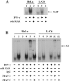
(A) Formation of GAF complexes in IFN-stimulated parental and L-C6 cell lines. Results for HeLa and L-C6 cells not treated (−) or treated (+) with IFN-γ are shown. Total cell lysates were analyzed by EMSA using a γ-32P-labeled hSIEm67 probe. The formation of GAF-specific transcription complexes was verified by incubating the IFN-treated cells with an excess nonradiolabeled hSIEm67 oligonucleotide. The arrow indicates the position of GAF complex. (B) Supershift analysis of GAF complexes, using STAT antibodies. IFN-γ-treated (+) or nontreated (−) HeLa and L-C6 cell extracts were incubated with STAT 1 and STAT 3 antibodies prior to incubation with γ-32P-labeled hSIEm67 probe and GAF complexes were analyzed by EMSA as mentioned for Fig. 3A. The arrow indicates the presence of the supershifted (S.S) GAF complexes. IgG, immunoglobulin G.
FIG. 4.
FIG. 4.
Inhibition of pY-STAT 1 levels in L-C6 cells. (A) Total cell extracts from HeLa and L-C6 cells not treated (−) or treated with IFN-γ (1,000 U/ml) at the indicated time intervals were subjected to SDS-PAGE followed by Western blot analysis with anti-pY-STAT 1 antibody (top), anti-STAT 1 antibody (middle), or antiactin antibody (bottom). (B) A similar experiment was performed as described for panel A, except that HeLa and L-C6 cells were treated with IFN-α.

References

    1. Bose, S., and A. K. Banerjee. 2003. Innate immune response against nonsegmented negative strand RNA viruses. J. Interferon Cytokine Res. 3:401-412. - PubMed
    1. Collins, P. L., R. M. Chanock, and K. McIntosh. 1996. Parainfluenza viruses, p. 1205-1241. In B. N. Fields, D. M. Knipe, and P. M. Howley (ed.), Fields virology, 3rd ed. Lippincott-Raven Publishers, Philadelphia, Pa.
    1. Delenda, C., G. Taylor, S. Hausmann, D. Garcin, and D. Kolakofsky. 1998. Sendai viruses with altered P, V, and W protein expression. Virology 242:327-337. - PubMed
    1. Durbin, A. P., J. M. McAuliffe, P. L. Collins, and B. R. Murphy. 1999. Mutations in the C, D and V open reading frames of human parainfluenza virus type 3 attenuate replication in rodents and primates. Virology 261:319-330. - PubMed
    1. Escoffier, C., S. Manie, S. Vincent, C. P. Muller, M. Billeter, and D. Gerlier. 1999. Nonstructural C protein is required for efficient measles virus replication in human peripheral blood cells. J. Virol. 73:1695-1698. - PMC - PubMed

Publication types

MeSH terms