. 2005 Jun;79(12):7883-8.
doi: 10.1128/JVI.79.12.7883-7888.2005.

Disruption of human TRIM5alpha antiviral activity by nonhuman primate orthologues

Affiliations

Disruption of human TRIM5alpha antiviral activity by nonhuman primate orthologues

Lionel Berthoux et al. J Virol. 2005 Jun.

Abstract

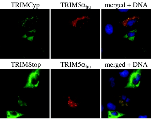
TRIM5 is a determinant of species-specific differences in susceptibility to infection by retroviruses bearing particular capsids. Human immunodeficiency virus type 1 (HIV-1) infection is blocked by the alpha isoform of macaque TRIM5 (TRIM5alpha(rh)) or by the product of the owl monkey TRIM5-cyclophilin A gene fusion (TRIMCyp). Human TRIM5alpha potently restricts specific strains of murine leukemia virus (N-MLV) but has only a modest effect on HIV-1. The amino termini of TRIM5 orthologues are highly conserved and possess a coiled-coil domain that promotes homomultimerization. Here we show that heterologous expression of TRIM5alpha(rh) or TRIMCyp in human cells interferes with the anti-N-MLV activity of endogenous human TRIM5alpha (TRIM5alpha(hu)). Deletion of the cyclophilin domain from TRIMCyp has no effect on heteromultimerization or colocalization with TRIM5alpha(hu) but prevents interference with anti-N-MLV activity. These data demonstrate that TRIM5 orthologues form heteromultimers and indicate that C-terminal extensions alter virus recognition by multimers of these proteins.


Figures

FIG. 1.
FIG. 1.
Inhibition of HIV-1 in human TE671 cells expressing owl monkey TRIMCyp or Rhesus macaque TRIM5α. LPCX-based retroviral vectors were used to transduce the indicated genes into human TE671 cells. Vect, Cyp, TS, TC and T5rh designate, respectively, Vector control, owl monkey CypA, TRIMStop, TRIMCyp, and TRIM5αrh. These cells were then infected for 16 h with HIV-1NL-GFP (pseudotyped vesicular stomatitis virus G protein) in the presence (+) or absence (-) of CsA (5 μM). One twentieth of the cells were maintained in culture for another day and used to determine the percentage of GFP-positive cells by FACS (bottom of the figure). Total DNA was extracted from the remainder of the cells 16 h after infection, and 5 μg of each DNA sample was analyzed by Southern blotting. The positions of the linear, 1-LTR, and 2-LTR HIV-1 cDNA species are indicated on the left. “Total” DNA refers to a band specific to all HIV-1 cDNA forms, including the integrated DNA.
FIG. 2.
FIG. 2.
TRIMCyp and TRIM5αrh abrogate the antiviral activity of endogenous TRIM5αhu. (A) TE671 cells expressing the indicated LPCX-derived constructs were infected with GFP-expressing B-MLV and N-MLV vectors at multiple doses. Cells were infected in the presence or absence of As2O3 (3 μM). Two days later, the percentage of infected cells was determined by FACS. (B) OMK cells were transduced with MIG (Vect) or with MIG-TRIM5αhu (T5hu) and challenged with DsRed-expressing N-MLV or B-MLV vectors. Two days later, the percentage of infected cells was determined by FACS. (C) OMK/MIG and OMK/MIG-T5hu cells were challenged with HIV-1-derived, DsRed-expressing CSRW vectors in the presence or absence of Cyclosporine A (2.5 μM).
FIG. 3.
FIG. 3.
Effect of TRIMStop on TRIM5αhu antiviral activity. (A) TE671-Vector (Vector) cells and TE671-TRIMStop cells were infected with HIV-1NL-GFP (left panel) or with N- and B-MLV (right panel). Two days later, the percentage of infected (GFP-expressing) cells was determined by FACS. (B) Total RNA was prepared from TE671-Vector (Vect), TE671-TRIMCyp (TC), and TE671-TRIMStop (TS), and RT-PCR was used to detect endogenous TRIM5α or TRIMCyp/TRIMStop. RT was performed in the presence (+) or absence (-) of reverse transcriptase. (C) 293T cells were transfected with LPCX (Vect), LPCX-TRIMCyp (TC), or LPCX-TRIMStop (TS), and Western blotting was performed using an antibody specific to TRIM5.
FIG. 4.
FIG. 4.
TRIMCyp and TRIMStop both bind TRIM5αhu. (A) Yeast two-hybrid system. TRIM5αhu, TRIM5αrh, TRIMCyp, TRIMStop, huTRIMStop, and HIV-1 Gag were fused to lexA. TRIMCyp and TRIMStop were expressed in fusion with the B42 activation domain. Pairs of fused LexA and B42 expression plasmids were transformed into Saccharomyces cerevisiae strain EGY48. For each transformant, β-galactosidase activity for three colonies is reported in Miller units with the standard deviation. (B) Binding in mammalian cells. TRIM5αhu was fused to GST. 293T cells were transfected with GST or GST-TRIM5αhu (GST-T5α) and cotransfected with LPCX (C), LPCX-TRIMCyp (TC), or LPCX-TRIMStop (TS). Thirty-six hours later, the cells were lysed in 50 mM Tris-Cl (pH 8.0), 150 mM NaCl, 1% NP40, 0.1% SDS, and GST was pulled down using glutathione-coated Sepharose beads (Pharmacia). One percent of the pre-pull-down lysate and 25% of the bound proteins were analyzed by Western blotting, using polyclonal antibodies directed against TRIM5, cyclophilin A, or GST.
FIG. 5.
FIG. 5.
Colocalization of TRIMCyp and TRIMStop with TRIM5αhu. TE671 cells were cotransfected with GST-TRIM5αhu and with 3× FLAG N-terminal-tagged versions of either TRIMCyp or TRIMStop. Thirty-six hours later, cells were fixed with 4% formaldehyde, permeabilized with 0.1% Triton X-100, and probed with antibodies against GST (rabbit polyclonal; Chemicon International) and FLAG (mouse monoclonal; Sigma). Fluorescent staining was done using Alexa488-conjugated goat anti-mouse and Alexa594-conjugated goat anti-rabbit antibodies and Hoechst33342 (all from Molecular Probes) to reveal DNA. Pictures were generated using a Nikon TE300 microscope with the Openlab 3.0 software.

Similar articles

Cited by

References

    1. Berthoux, L., S. Sebastian, E. Sokolskaja, and J. Luban. 2004. Lv1 inhibition of human immunodeficiency virus type 1 is counteracted by factors that stimulate synthesis or nuclear translocation of viral cDNA. J. Virol. 78:11739-11750. - PMC - PubMed
    1. Berthoux, L., G. J. Towers, C. Gurer, P. Salomoni, P. P. Pandolfi, and J. Luban. 2003. As2O3 enhances retroviral reverse transcription and counteracts Ref1 antiviral activity. J. Virol. 77:3167-3180. - PMC - PubMed
    1. Bieniasz, P. D. 2003. Restriction factors: a defense against retroviral infection. Trends Microbiol. 11:286-291. - PubMed
    1. Braaten, D., C. Aberham, E. K. Franke, L. Yin, W. Phares, and J. Luban. 1996. Cyclosporine A-resistant human immunodeficiency virus type 1 mutants demonstrate that Gag encodes the functional target of cyclophilin A. J. Virol. 70:5170-5176. - PMC - PubMed
    1. Colgan, J., H. E. Yuan, E. K. Franke, and J. Luban. 1996. Binding of the human immunodeficiency virus type 1 Gag polyprotein to cyclophilin A is mediated by the central region of capsid and requires Gag dimerization. J. Virol. 70:4299-4310. - PMC - PubMed

Publication types

MeSH terms

LinkOut - more resources