. 2005 Jun;166(6):1585-92.
doi: 10.1016/S0002-9440(10)62469-8.

Mechanism of acute fetal cardiovascular depression after maternal inflammatory challenge in mouse

Affiliations

Mechanism of acute fetal cardiovascular depression after maternal inflammatory challenge in mouse

Samuli Rounioja et al. Am J Pathol. 2005 Jun.

Abstract

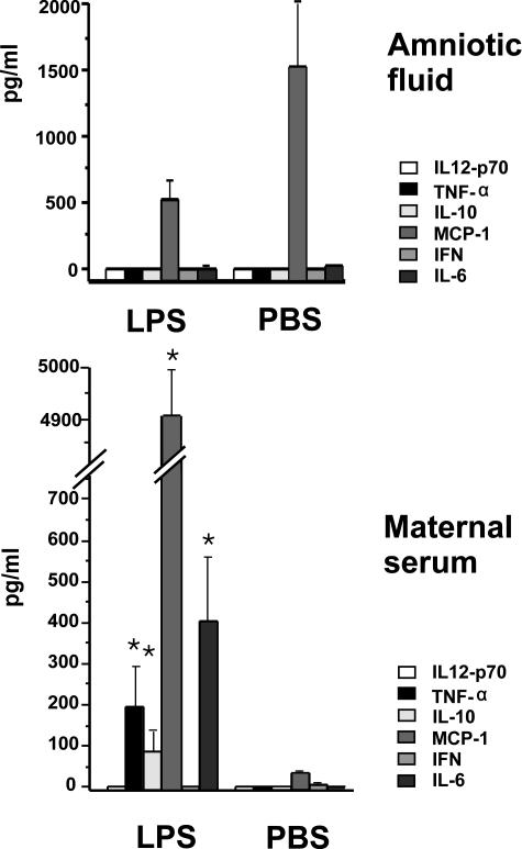
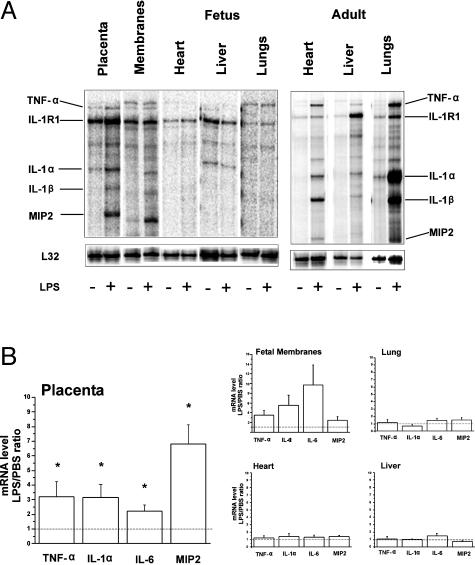
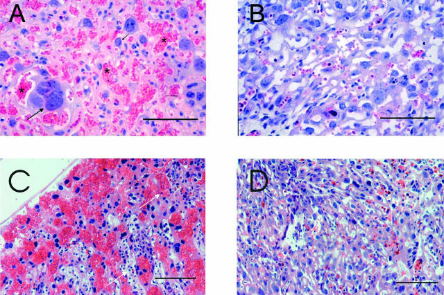
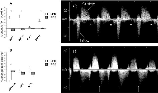
Intra-amniotic lipopolysaccharide (LPS) causes an acute inflammatory response and cardiac dysfunction in fetal mice. We hypothesized that the placenta protects the fetus against maternally administered bacterial toxins, delaying the onset of a fetal inflammatory response and vascular compromise. At 14 to 15 days of gestation, DBA mice were randomized to receive LPS (2.4 mg/kg) or vehicle intraperitoneally. Doppler ultrasonography of fetal cardiovascular hemodynamics was performed before and 6 hours after maternal LPS. Six hours after the LPS, maternal serum concentrations of tumor necrosis factor-alpha and interleukin (IL)-6 (P < 0.05) were increased. Placenta showed severe maternal vascular dilatation and congestion. The expressions of tumor necrosis factor-alpha, IL-1alpha, and IL-6 (P < 0.05) were increased, and the expression of Toll-like receptor 4 was constitutive in placenta. The expression of Toll-like receptor 2 increased (P < 0.05) and was detected in labyrinthine macrophages. No inflammatory activation was found in fetal tissues, and amniotic fluid revealed no significant increase in cytokines. The ultrasonographic examination demonstrated increased fetal cardiac afterload after LPS, with 65% of the fetuses exhibiting atrioventricular valve regurgitation. In conclusion, maternal inflammatory insult activates placental labyrinthine macrophages leading to an acute increase in placental vascular resistance and fetal cardiac dysfunction without an inflammatory response in fetus.


Figures

Figure 1
Figure 1
Concentrations of inflammatory mediators in maternal serum and amniotic fluid 6 hours after intraperitoneal LPS (2.4 mg/kg) or vehicle. Results are expressed as mean ± SEM.
Figure 2
Figure 2
Expressions of inflammatory mediators 6 hours after maternal LPS (2.4 mg/kg) or vehicle. A: Representative gel electrophoresis of multiple mRNAs in fetal and maternal tissues by ribonuclease protection assay. B: Expressions of inflammatory mediators in fetal and gestational tissues. The data are expressed as a fold increase in mRNA levels in the LPS group relative to the vehicle group. The results are mean ± SEM (n = 5 to 12 animals per group). *P < 0.05 versus vehicle group. Only the mediators induced by LPS in the placenta are shown.
Figure 3
Figure 3
Histopathology of placenta by Leder’s staining. A: Six hours after intraperitoneal LPS administration, severe vascular congestion in the labyrinthine part of the placenta is seen. Cytotrophoblast (black arrow) and maternal red blood cells as shown with asterisk. B: Placenta 6 hours after vehicle administration (PBS) shows no congestion. Placenta 12 hours after LPS (C) and PBS (D). Note the accumulation of lymphocytes and granulocytes (white arrow) after LPS. Scale bars, 50 μm.
Figure 4
Figure 4
A: Immunostaining of mouse placenta. Highly specific positive staining for TLR2 antibody appears in red in placental macrophages in labyrinthine mesenchyme. B: Control staining without secondary antibody. Bottom: The mRNA expression of TLR2 and TLR4 in placenta 6 hours after intraperitoneal LPS and vehicle. The bars indicate the fold increase compared to vehicle-treated animals. Results are mean ± SEM.
Figure 5
Figure 5
Left: Ultrasonographic parameters of fetal circulation (A) and cardiac function (B) 6 hours after LPS or vehicle (PBS) administration. Data are presented as change (%) from the baseline values. Right: C: Inflow and outflow blood velocity waveforms of the fetal heart obtained 6 hours after vehicle, semilunar valve closure is shown as a valve click (*). D: After LPS administration, semilunar valve regurgitation is seen. The valve click is missing, and regurgitation starts immediately after the end of ejection (arrow). PI, pulsatility index; PIV, pulsatility index for veins; UA, umbilical artery; DAO, descending aorta; ICA, intracranial artery; DV, ductus venosus; OFVmean, outflow mean velocity; IRT, isovolumetric relaxation time; ICT, isovolumetric contraction time. Data are expressed as mean ± SEM.

Similar articles

Cited by

References

    1. Natanson C, Eichenholz PW, Danner RL, Eichacker PQ, Hoffman WD, Kuo GC, Banks SM, MacVittie TJ, Parrillo JE. Endotoxin and tumor necrosis factor challenges in dogs simulate the cardiovascular profile of human septic shock. J Exp Med. 1989;169:823–832. - PMC - PubMed
    1. Rangel-Frausto MS, Pittet D, Costigan M, Hwang T, Davis CS, Wenzel RP. The natural history of the systemic inflammatory response syndrome (SIRS). A prospective study. JAMA. 1995;273:117–123. - PubMed
    1. Parrillo JE, Parker MM, Natanson C, Suffredini AF, Danner RL, Cunnion RE, Ognibene FP. Septic shock in humans. Advances in the understanding of pathogenesis, cardiovascular dysfunction, and therapy. Ann Intern Med. 1990;113:227–242. - PubMed
    1. Beutler B. Endotoxin, Toll-like receptor 4, and the afferent limb of innate immunity. Curr Opin Microbiol. 2000;3:23–28. - PubMed
    1. Tak PP, Firestein GS. NF-kappaB: a key role in inflammatory diseases. J Clin Invest. 2001;107:7–11. - PMC - PubMed

Publication types

MeSH terms