Comparative Study
. 2005 Jun;166(6):1593-606.
doi: 10.1016/S0002-9440(10)62470-4.

Gender-based differences in bleomycin-induced pulmonary fibrosis

Affiliations
Comparative Study

Gender-based differences in bleomycin-induced pulmonary fibrosis

Mehrnaz Gharaee-Kermani et al. Am J Pathol. 2005 Jun.

Abstract

The role of gender and sex hormones is unclear in host response to lung injury, inflammation, and fibrosis. To examine gender influence on pulmonary fibrosis, male and female rats were given endotracheal injections of either saline or bleomycin. Female rats showed higher mortality rates and more severe fibrosis than did male rats, as indicated by higher levels of lung collagen deposition and fibrogenic cytokine expression. To clarify the potential role of female sex hormones in lung fibrosis, female rats were ovariectomized and treated with either estradiol or vehicle plus endotracheal injections of either saline or bleomycin. The results showed diminished fibrosis in the ovariectomized, bleomycin-treated rats without hormone replacement. Estradiol replacement restored the fibrotic response to that of the intact female mice in terms of lung collagen deposition and cytokine expression, which was accompanied by higher plasma estradiol levels. Furthermore, fibroblasts from bleomycin-treated rats exhibited increased responsiveness to estradiol treatment, causing dose-dependent increases in procollagen 1 and transforming growth factor-beta1 mRNA expression relative to untreated controls. Taken together these findings suggest that female mice may have an exaggerated response to lung injury relative to male mice because of female sex hormones, which have direct fibrogenic activity on lung fibroblasts. This may provide a mechanism for a hormonally mediated intensification of pulmonary fibrosis.


Figures
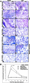
Figure 1
Figure 1
Effects of gender on lung histopathology eosinophil recruitment. Representative chromotrope 2R-stained lung section from day 7, 14, and 21 female (a, c, e) and male (b, d, f) after BLM treatment and corresponding control at day 21 is shown (g, h). At day 7 after BLM, treated female lung sections show greater inflammatory cell infiltration and eosinophil (arrows and inset showing higher magnification) recruitment (a) compared to those in male lungs (b). Fibrotic lesions became more extensive at days 14 and 21 after BLM in female (c, e) than that observed in male rats (d, f). Saline-treated controls showed no sign of inflammation in both genders at the day 21 time point (g, h). A higher magnification view of the cellular areas shows dense clusters of polymorphonuclear cells that stained red with the chromotrope 2R (inset in a). Eosinophils in lung section were counted as described in Materials and Methods and the results are shown in B as the number of cells per high-power field. The values were presented as means ± SE (n = 4 per group). The increases in the number of eosinophils in female relative to respective male in BLM-treated animals were statistically significant (P < 0.001) for day 7. Masson trichrome-stained lung sections revealed greater amounts of collagen deposition in female lungs at days 14 and 21 after BLM treatment compared to males (i–l). In contrast, SAL controls corresponding to female and male rats did not show any abnormal collagen staining (m, n). Original magnification, ×400 (inset in a).
Figure 2
Figure 2
Effects of gender on lung hydroxyproline content. PF was biochemically assessed by measurement of total lung hydroxyproline content on day 21 after BLM treatment. Results in BLM-treated lungs were expressed as a percentage of the corresponding SAL-treated controls, and shown as means ± SE (n = 10) for both female and male experimental groups. Lung hydroxyproline content was significantly higher in BLM-treated female versus male rats (P < 0.005). The lung hydroxyproline contents of male and female SAL-treated control rats were not significantly different.
Figure 3
Figure 3
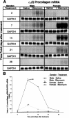
Effect of gender on collagen gene expression. Total lung RNA from control and BLM-treated female and male animals at indicated time points were subjected to Northern blot analysis for determination of procollagen α1 (I) mRNA as described in Materials and Methods. A: Autoradiographs for collagen mRNA and GAPDH are shown. GAPDH mRNA was used to confirm uniform RNA loading and for use as a normalization factor. Each lane represents a RNA sample from a single animal. B: Bands on the autoradiographs were quantitated by an Ambis radioactive-imaging system. The data represent means ± SE of results from three animals at each time point. The increases in procollagen mRNA in female relative to male lungs were statistically significant (P < 0.01) for days 7 and 14 after BLM treatment.
Figure 4
Figure 4
Lung cytokine mRNA expression. Total lung RNA from control and BLM-treated female and male rats at the indicated time points was subjected to Northern blot for determination of TGF-β1, MCP-1, and TNF-α mRNAs as described in Materials and Methods. The autoradiographs for TGF-β1 and GAPDH mRNAs are shown in A, with the GAPDH mRNA level being used to document uniform RNA loading and to normalize the cytokine mRNA results. The results of direct quantitation using an Ambis radioactivity imaging system for TGF-β1, MCP-1, and TNF-α mRNA levels are shown in B, C, and D, respectively. Each lane represented an RNA sample from a single animal and the results are shown as means ± SE of three animals at each time point. B: The levels of TGF-β1 mRNA in day 7 BLM-treated female rats were significantly (P < 0.001) higher than those in male rats. C: The increases in MCP-1 mRNA in females relative to respective male were statistically significant (P < 0.001) for days 3, 7, and 14 after BLM treatment. D: TNF-α mRNA was also significantly higher in female versus male lungs (P < 0.01) on day 3 after BLM treatment.
Figure 5
Figure 5
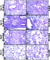
The effects of ovarian dysfunction on lung histology. H&E-stained lung sections from each treatment group are shown. A: OV + BLM; B: N + BLM; C: OV + E + BLM; D: OV + V + BLM; E: OV + V + SAL; F: N + V + SAL. Lung sections from ovariectomized rats on day 14 after BLM (A) showed a lower numbers of monocytes, macrophages, and eosinophils and less fibrosis compared with corresponding sham-operated group (B). This diminution was overcome by restoration of estradiol levels in ovariectomized BLM-treated group (C, E) relative to corresponding control (D, F). In contrast, control SAL-treated lungs from ovariectomized or normal received vehicle rats did not demonstrate any evidence of inflammation (G and H, respectively). N, normal sham operation; E, 17β-estradiol; V, vehicle.
Figure 6
Figure 6
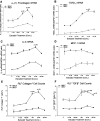
Effects on lung collagen and plasma estradiol. The effects of OV with or without estradiol replacement therapy are shown for lung hydroxyproline content on day 21 after BLM (A), lung α1(1) procollagen mRNA on day 14 after BLM (B), plasma estradiol level on day 21 after BLM (C), and comparison of plasma estradiol levels with lung hydroxyproline content in all groups (D). Lung hydroxyproline content, procollagen I mRNA, and plasma estradiol levels from normal and ovariectomized rats that received estradiol (E) or vehicle (V) and treated with BLM or SAL are shown as means ± SE (n = 5). A: Lung hydroxyproline content was significantly higher in ovariectomized and sham-operated rats that received estradiol and BLM (E/BLM) than in ovariectomized rats that received vehicle and BLM (V/BLM). Whole-lung RNA was isolated from all groups of rats at day 14 and analyzed for α1(1) procollagen and GAPDH (for normalization) mRNA by RT-PCR as described in Materials and Methods. The results were normalized to the GAPDH mRNA signal and expressed as the ratio of α1(1) procollagen to GAPDH mRNA signals. Means ± SE from three animals in each group are shown. B: Significantly higher α1(1) procollagen mRNA levels were seen as a result of BLM and estradiol treatment (E/BLM) in ovariectomized and normal rats compared to that in ovariectomized rats that received vehicle (V/BLM). Three weeks after BLM challenge, blood was collected and plasma estradiol levels measured by enzyme-linked immunosorbent assay. C: Results showed a significant decrease in plasma estradiol levels at 3 weeks after OV (V/BLM) with return to normal levels after estradiol (E/BLM) replacement therapy. D: Linear regression analysis was attempted to see if plasma estradiol levels would correlate with lung hydroxyproline content. The regression line (r = 0.405, P = 0.029) is shown with boundary lines indicating the 95% confidence intervals. Although the correlation was relatively weak, it was statistically significant.
Figure 7
Figure 7
The effects of ovarian dysfunction on BLM lung cytokine expression. Lung TGF-β1 (A), IL-4 (B), MCP-1 (C), and IFN-γ (D) mRNAs were examined by RT-PCR analysis as described in the legend to Figure 6. A, B: At day 14 after BLM treatment the levels of TGF-β1 (P < 0.001) and IL-4 (P < 0.02) mRNAs were significantly higher as a result of estradiol treatment (E/BLM) in both ovariectomized and normal rats compared to those in ovariectomized rats receiving vehicle (V/BLM) only. C: However the expression of MCP-1 was not significantly affected by estradiol treatment. D: Whereas the expression of IFN-γ mRNA was significantly lower (P < 0.002) in ovariectomized rats receiving estradiol (E/BLM) and in normal rats receiving vehicle (V/BLM) relative to that in ovariectomized rats treated with vehicle (V/BLM). Estradiol had no significant effect in normal rats (V/BLM versus E/BLM).
Figure 8
Figure 8
Effects of estradiol on fibrotic lung fibroblasts in vitro. Rat lung fibroblasts from BLM -treated (BRLF) and normal (NRLF) rats were isolated and treated with various concentration of estradiol. The cells were harvested for analysis of procollagen α1 (I) (A), TGF-β1 (B), IL-4 (C), and MCP-1 (D) mRNAs by RT-PCR. The procollagen, TGF-β1, and IL-4 mRNAs gradually increased after treatment with increasing doses of estradiol, with significant increases at concentrations of 1 and 10 nmol/L (only at 10 nmol/L for IL-4) in BRLF compared untreated control BRLF. D: The expression of MCP-1 was not significantly affected by estradiol treatment. Collagen type 1 and TGF-β1 expression was also assessed by immunohistochemistry and expressed as a percentage of total cells positively stained by the respective anti-collagen (E) or TGF-β1 (F) antibodies.

Similar articles

Cited by

References

    1. Hiwatari N, Shimura S, Sasaki T, Aikawa T, Ando Y, Ishihara H, Seskizawa K, Sasakima H, Takishima T. Prognosis of idiopathic pulmonary fibrosis in patients with mucous hypersecretion. Am Rev Respir Dis. 1991;143:182–185. - PubMed
    1. Mannino DM, Etzel RA, Parrish RG. Pulmonary fibrosis deaths in the United States, 1979–1991; an analysis of multiple-cause mortality data. Am J Respir Crit Care Med. 1996;153:1548–1552. - PubMed
    1. Gross TJ, Hunninghake GW. Idiopathic pulmonary fibrosis. N Engl J Med. 2001;345:517–525. - PubMed
    1. Piguet PF, Collart MA, Grau GE, Sappino AP, Vassalli P. Requirement of tumor necrosis factor for development of silica-induced pulmonary fibrosis. Nature. 1990;344:245–247. - PubMed
    1. Gharaee-Kermani M, Phan SH. Role of cytokines and cytokine therapy in wound healing and fibrotic diseases. Curr Pharm Dis. 2001;7:1083–1103. - PubMed

Publication types

MeSH terms