. 2005 Jun;166(6):1781-91.
doi: 10.1016/S0002-9440(10)62488-1.

Teratoma formation leads to failure of treatment for type I diabetes using embryonic stem cell-derived insulin-producing cells

Affiliations

Teratoma formation leads to failure of treatment for type I diabetes using embryonic stem cell-derived insulin-producing cells

Takahisa Fujikawa et al. Am J Pathol. 2005 Jun.

Abstract

Embryonic stem (ES) cells have been proposed to be a powerful tool in the study of pancreatic disease, as well as a potential source for cell replacement therapy in the treatment of diabetes. However, data demonstrating the feasibility of using pancreatic islet-like cells differentiated from ES cells remain controversial. In this study we characterized ES cell-derived insulin-expressing cells and assessed their suitability for the treatment of type I diabetes. ES cell-derived insulin-stained cell clusters expressed insulin mRNA and transcription factors associated with pancreatic development. The majority of insulin-positive cells in the clusters also showed immunoreactivity for C-peptide. Insulin was stored in the cytoplasm and released into the culture medium in a glucose-dependent manner. When the cultured cells were transplanted into diabetic mice, they reversed the hyperglycemic state for approximately 3 weeks, but the rescue failed due to immature teratoma formation. Our studies demonstrate that reversal of hyperglycemia by transplantation of ES cell-derived insulin-producing cells is possible. However, the risk of teratoma formation would need to be eliminated before ES cell-based therapies for the treatment of diabetes are considered.


Figures

Figure 1
Figure 1
ES cells differentiated into pancreatic islet-like cell clusters via in vitro culture system. A: Dense clusters of cells appeared after 7 to 10 days of the differentiation culture step and they proliferated and grew during the following periods. B: By the end of this stage the number of nestin-positive cells significantly decreased and islet-like clusters were often expanded in number and became the most predominant cell type with up to 40% of the cultures. C: Most of cells among the clusters expressed insulin (red) by immunohistochemistry (blue, DAPI). D: Of the cells in the clusters, 86.1 ± 2.1% were insulin-positive by flow cytometry (green area, clusters before transplantation; dotted curve, negative control). Scale bars, 100 μm.
Figure 2
Figure 2
Pancreatic gene expression in undifferentiated ES cells and ES cell-derived cell clusters. A and B: RT-PCR analyses for expression of Glut-2 and pancreatic endocrine proteins and transcription factors. Panc, normal mouse pancreas tissue. A: Glut-2 gene and other endocrine pancreatic mRNAs were expressed in the ES cell-derived differentiated cell clusters. B: The cell clusters also expressed messages of all of the transcriptional factors found in committed endocrine and pancreatic lineages, but not OCT-4. C: Northern blot of the cell clusters to examine the expression of insulin messages. RNAs extracted from INS-1 cells and β-TC-6 cells were used as positive controls. Those from WB-F344 cells were used as a negative control. The results show the presence of both insulin I and II mRNAs at quantitatively sufficient amounts in cultured ES-derived cell clusters.
Figure 3
Figure 3
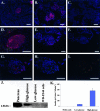
A to I: Insulin and C-peptide immunohistochemistry analyses using ES cell-derived islet-like cell clusters. A–F: Immunohistochemistry using each single antibody: insulin staining (red) with DAPI nuclear staining (blue) (A–C), C-peptide staining (red) with DAPI (blue) (D–F). A, D: Tissues from normal mouse pancreas were used as positive control. C, F: Negative controls were obtained from the samples without primary antibodies. B, E: The ES cell-derived clusters did express both insulin and C-peptide proteins ubiquitously in the cytoplasms. G–I: Double staining using both insulin (red) and C-peptide (green) antibodies (blue, DAPI). G: Merged image of insulin and DAPI, H: C-peptide and DAPI, and I: insulin and C-peptide. Yellow-colored cells indicate insulin and C-peptide double-positive cells. Most cells in the clusters showed co-expression of C-peptide and mature insulin. J: Western blot analysis of insulin storage after collection of cell lysates after a 2-hour culture using low (5.5 mmol/L)- or high (25 mmol/L)-glucose conditions. The differentiated ES cell-derived cell clusters synthesize and store sufficient amounts of insulin during exposure to high-glucose media, whereas only small amounts of insulin were detected in the cell lysate from low-glucose media. K: ELISA assay for insulin in conditioned media from ES cell-derived cell clusters exposed to low- or high-glucose conditions for 2 hours. Challenge of ES cell-derived cell clusters (∼90 to 100 clusters per experiment) with high-glucose concentration resulted in secretion of 283.1 ± 41.2 μU.islets−1.60 minutes−1 of insulin into the media, which is significantly higher than that in low-glucose media (35.2 ± 2.2 μU.islets−1.60 minutes−1, P < 0.05) or in media alone (0 μU.islets−1.60 minutes−1, P < 0.05). Scale bars, 100 μm.
Figure 4
Figure 4
Transplantation of ES cell-derived insulin-producing cell clusters into STZ-treated diabetic NOD/scid mice. Shown in the graph are control STZ-treated mice with no cell transplantation (dotted line, n = 5) and mice transplanted with 100 to 150 ES cell-derived insulin-producing cell clusters into the renal subcapsular space (bold solid line and solid line, n = 10). Control mice exhibited persistent hyperglycemia and all died by day 20. The mice receiving the transplant began to reverse their high blood glucose levels within 2 to 3 days and these levels were maintained up to 3 weeks after transplantation. Two of ten mice died without hyperglycemia (solid line), whereas all other mice eventually became hyperglycemic and died by day 30 (bold solid line, n = 8).
Figure 5
Figure 5
Analysis of graft after kidney excision via H&E staining and immunohistochemistry and RT-PCR for insulin expression. A: Macroscopically, an encapsulated solid tumor was found at each transplanted site in 6 of 10 transplanted mice. B, C: H&E staining of tumor shows immature epithelial cells including mature gland tissues (B, arrows), foci of squamous epithelial cells (B, circle), and immature muscle in the background stroma (C, arrows), which confirmed it as teratoma. D, E: Insulin immunohistochemistry of the graft tissues (green, GFP). E is a highly magnified image of the boxed area in D. Very small numbers of the cells in the tumor were positive for insulin (red). F: RT-PCR analysis of the graft tissues for expression of insulin I and II. Both insulin I and II gene expressions were detected but down-regulated dramatically after transplantation. Scale bars, 100 μm.
Figure 6
Figure 6
Analysis of OCT-4 and SSEA-1 expressions before and after transplantation. A: By RT-PCR, the expression of OCT-4 was detected in the graft tissues even though the transplanted cells used had no OCT-4 expression before transplantation. B: By flow cytometry, 0.23 ± 0.03% of the cells in clusters were SSEA-1-positive (green area, clusters before transplantation; dotted curve, negative control; solid curve with no filling, positive control using undifferentiated ES cells). C–H: OCT-4 and SSEA-1 immunohistochemistry analyses. C–E: OCT-4 staining (green) with DAPI (blue). F–H: SSEA-1 staining (green) with DAPI (blue). C, F: Undifferentiated ES cells were used as positive control. D, G: Neither OCT-4 nor SSEA-1 were positive in the clusters before transplantation. E, H: Some of the colonized OCT-4- and SSEA-1-positive cells were observed in the whole grafted tissues after transplantation. Scale bars, 100 μm.

References

    1. Sutherland DE, Gruessner RW, Dunn DL, Matas AJ, Humar A, Kandaswamy R, Mauer SM, Kennedy WR, Goetz FC, Robertson RP, Gruessner AC, Najarian JS. Lessons learned from more than 1,000 pancreas transplants at a single institution. Ann Surg. 2001;233:463–501. - PMC - PubMed
    1. Ricordi C, Strom TB. Clinical islet transplantation: advances and immunological challenges. Nat Rev Immunol. 2004;4:259–268. - PubMed
    1. Lechner A, Leech CA, Abraham EJ, Nolan AL, Habener JF. Nestin-positive progenitor cells derived from adult human pancreatic islets of Langerhans contain side population (SP) cells defined by expression of the ABCG2 (BCRP1) ATP-binding cassette transporter. Biochem Biophys Res Commun. 2002;293:670–674. - PubMed
    1. Suzuki A, Nakauchi H, Taniguchi H. Prospective isolation of multipotent pancreatic progenitors using flow-cytometric cell sorting. Diabetes. 2004;53:2143–2152. - PubMed
    1. Shapiro AM, Lakey JR, Ryan EA, Korbutt GS, Toth E, Warnock GL, Kneteman NM, Rajotte RV. Islet transplantation in seven patients with type 1 diabetes mellitus using a glucocorticoid-free immunosuppressive regimen. N Engl J Med. 2000;343:230–238. - PubMed

Publication types