Comparative Study
. 2005 Jun;166(6):1793-806.
doi: 10.1016/S0002-9440(10)62489-3.

Dual infection with Helicobacter bilis and Helicobacter hepaticus in p-glycoprotein-deficient mdr1a-/- mice results in colitis that progresses to dysplasia

Affiliations
Comparative Study

Dual infection with Helicobacter bilis and Helicobacter hepaticus in p-glycoprotein-deficient mdr1a-/- mice results in colitis that progresses to dysplasia

Lillian Maggio-Price et al. Am J Pathol. 2005 Jun.

Abstract

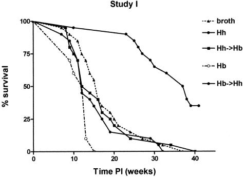
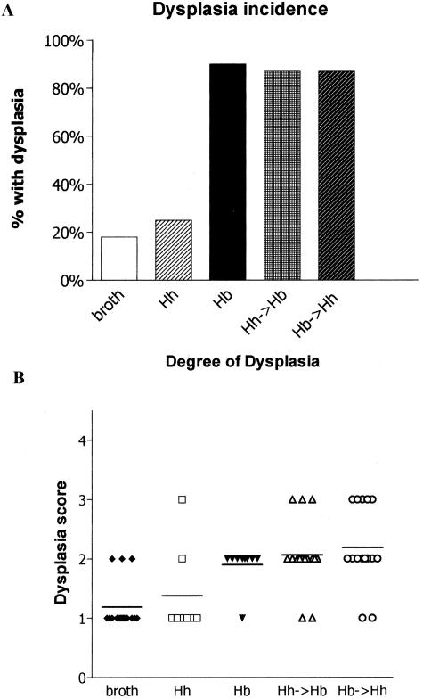
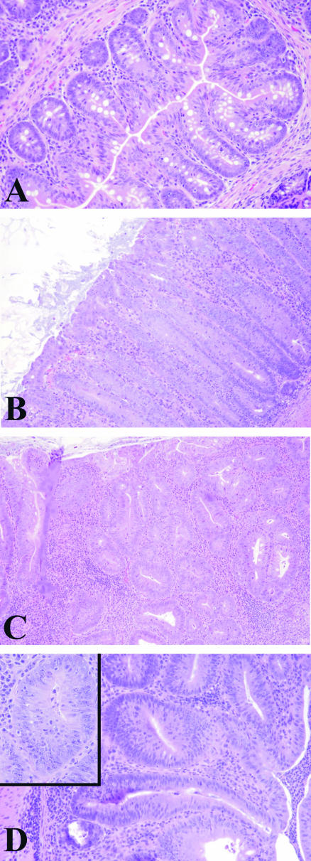
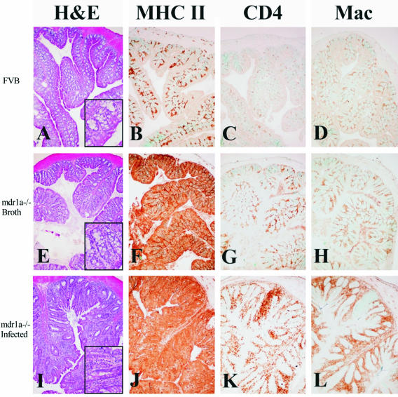
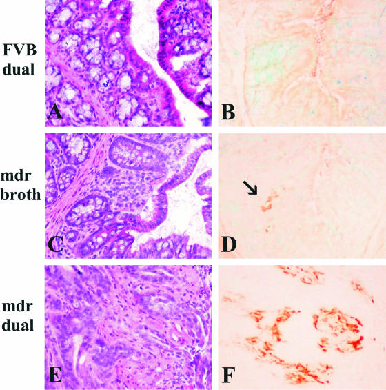
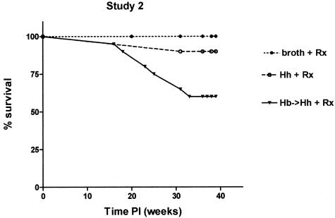
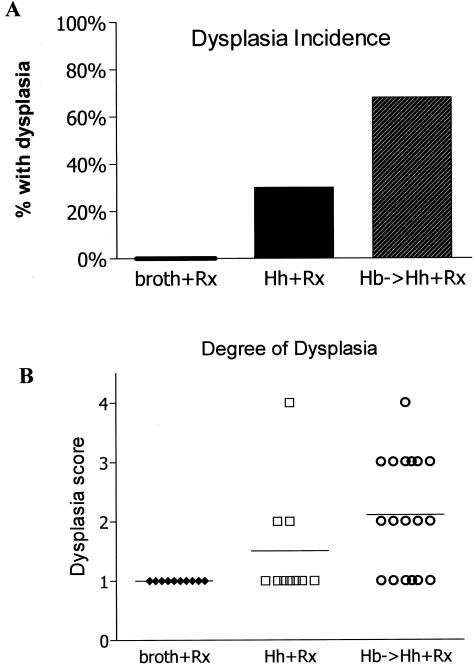
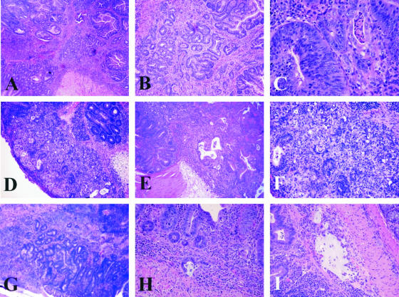
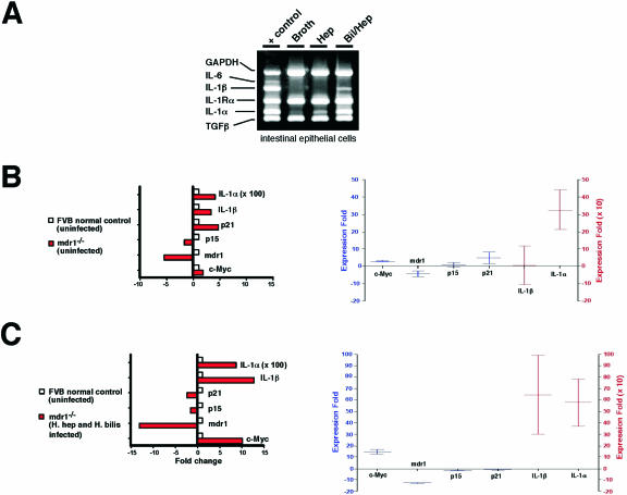
Patients with inflammatory bowel disease (IBD) are at increased risk for developing high-grade dysplasia and colorectal cancer. Animal IBD models that develop dysplasia and neoplasia may help elucidate the link between inflammation and colorectal cancer. Mdr1a-/- mice lack the membrane efflux pump p-glycoprotein and spontaneously develop IBD that can be modulated by infection with Helicobacter sp: H. bilis accelerates development of colitis while H. hepaticus delays disease. In this study, we determined if H. hepaticus infection could prevent H. bilis-induced colitis. Unexpectedly, a proportion of dual-infected mdr1a-/- mice showed IBD with foci of low- to high-grade dysplasia. A group of dual-infected mdr1a-/- animals were maintained long term (39 weeks) by intermittent feeding of medicated wafers to model chronic and relapsing disease. These mice showed a higher frequency of high-grade crypt dysplasia, including invasive adenocarcinoma, possibly because H. hepaticus, in delaying the development of colitis, allows time for transformation of epithelial cells. Colonic epithelial preparations from co-infected mice showed increased expression of c-myc (5- to 12-fold) and interleukin-1alpha/beta (600-fold) by real-time polymerase chain reaction relative to uninfected wild-type and mdr1a-/- animals. This animal model may have particular relevance to human IBD and colorectal cancer because certain human MDR1 polymorphisms have been linked to ulcerative colitis and increased risk for colorectal cancer.


Figures

Figure 1
Figure 1
Percent survival of Helicobacter-infected and uninfected mdr1a−/− mice. The addition of H. hepaticus to H. bilis did not increase survival. H. hepaticus-infected mdr1a−/− mice have significantly increased (P < 0.01) survival relative to all other groups.
Figure 2
Figure 2
Incidence and degree of dysplasia in uninfected and Helicobacter-infected mdr1a−/− mice (study 1). A: Infection of mdr1a−/− mice with H. bilis alone or both H. bilis and H. hepaticus had the highest incidence of dysplasia. B: Dual-infected mdr1a−/− mice exhibited the most severe dysplasia relative to uninfected mice (P < 0.001) or singly infected mice (P < 0.05); 1, no dysplasia; 2, low grade; 3, high grade; 4, high grade with invasion/adenocarcinoma).
Figure 3
Figure 3
H&E-stained paraffin-embedded sections of colons from mdr1a−/− mice demonstrating the spectrum of mucosal lesions observed in study 1. A: Relatively normal mucosa from a nonclinically colitic mdr1a−/− mouse. Note low-grade inflammation in the lamina propria confined to the mucosa. B: Hyperplastic colitis characterized by a diffuse moderately thickened mucosa with elongated glands, reduction in goblet cells, and increased mitotic figures. C: Low-grade dysplasia. Note irregularly branching glands, hyperchromatic nuclei, and loss of maturation gradient toward the lumen. D: High-grade dysplasia. Note irregular glands lined by crowded epithelial cells. The nuclei are pleomorphic, hyperchromatic, and stratified (inset). Original magnifications: ×10; ×40 (inset).
Figure 4
Figure 4
OCT-embedded frozen colon section with immunohistochemical detection of MHC class II (B, F, J), CD4+ cells (C, G, K), and F4/80+ macrophages (D, H, L) in the colon of FVB wild-type (B, C, D), broth control (F, G, H), and dual-infected (J, K, L) mdr1a−/− mice. In the normal, noninflamed colon of FVB mice (A), MHC class II expression is restricted to small numbers of leukocytes in the lamina propria (B) and numbers of CD4+ cells and macrophages are low (C, D). In contrast, there are subtle proliferative and inflammatory changes in broth-uninfected mdr1a−/− mice (E), which is accompanied by pronounced up-regulation of MHC class II expression in enterocytes (F) and moderately increased numbers of CD4+ cells and macrophages in lamina propria (G, H). In animals dual-infected with H. bilis and H. hepaticus, there are severe proliferative and inflammatory changes in the colon (I), accompanied by significant up-regulation of MHC class II on enterocytes and infiltrating leukocytes (J) and marked infiltration of CD4+ T cells (K) and macrophages (L) in lamina propria. Original magnifications, ×10.
Figure 5
Figure 5
OCT-embedded frozen colon sections from dual-infected FVB (A and B), uninfected mdr1a−/− (C and D), and dual-infected mdr1a−/− mice (E and F). B: Dual-infected FVB wild-type mice remain lesion-free and no Cox-2-positive cells are found in the colonic mucosa. D: Uninfected mdr1a−/− mice show rare Cox-2-positive cells (arrow) in the mild, spontaneous inflammatory infiltrates of the colon. F: In contrast, dual-infected mdr1a−/− mice have large numbers of cells in the lamina propria of colon (and cecum, data not shown) that express high levels of Cox-2. Note diffuse light background staining of intestinal contents, which is especially prominent in B and D. Original magnifications, ×20.
Figure 6
Figure 6
Percent survival of Helicobacter-infected and uninfected mdr1a−/− mice in study 2. Prolonged survival of uninfected and dual-infected (H. bilis and H. hepaticus) mdr1a−/− mice was achieved by feeding medicated wafers. Dual-infected mdr1a−/− mice had decreased survival (P < 0.05) relative to uninfected mdr1a−/− mice and exhibited a high incidence of dysplasia.
Figure 7
Figure 7
Incidence and degree of dysplasia in uninfected and Helicobacter-infected mdr1a−/− mice (study 2). Dual-infected mdr1a−/− mice had the highest incidence of dysplasia (A) and exhibited the most severe dysplasia relative to uninfected mice (P < 0.001) or singly infected mice (P < 0.05) (B). 1, no dysplasia; 2, low grade; 3, high grade; 4, high grade with invasion/adenocarcinoma.
Figure 8
Figure 8
Histopathology of high-grade dysplasia and invasive adenocarcinoma in dual-infected mdr1a−/− mice. A: Colonic high-grade dysplasia (grade 3). B: Higher power of A. C: Cecal high-grade dysplasia (grade 3). Note haphazard stratification of hyperchromatic nuclei. D: Cecal high-grade dysplasia/medullary carcinoma in situ. E: Deeper serial section of D. F: Higher magnification of D. G: Invasive colonic adenocarcinoma. Note irregular glands present at the submucosa junction muscularis externa. H: Invasive colonic mucinous adenocarcinoma. I: Additional section of H demonstrating lake of mucin within a dilated submucosal lymphatic. Original magnifications: ×4 (A, G); ×10 (B, D, E); ×20 (F, H, I); ×40 (C).
Figure 9
Figure 9
A: Multiplex PCR for multiple cytokines in colonic epithelial cells from uninfected and infected mdr1a−/− mice. Increased expression of IL-1α and IL-1β was only noted in colonic epithelial cells of mdr1a−/− mice infected with both H. bilis and H. hepaticus and not in epithelial cells from H. hepaticus or uninfected broth animals. Real-time PCR showing expression of c-myc and IL-1α and IL-1β in colonic epithelial cells from uninfected mdr1a−/− and FVB+/+ mice (B) and from uninfected FVB+/+ and dual-infected mdr1a−/− mice (C).
Figure 10
Figure 10
Real-time PCR for H. bilis and H. hepaticus from fecal samples from mdr1a−/− mice. A: H. bilis is the predominant organism in dual-infected animals in study 1. B: Medicated feed has reduced numbers of H. bilis in dual-infected animals in study 2. C: Medicated feed has dramatically reduced H. hepaticus in dual-infected animals in study 2. Fold-expression was generated by the CT method, whereby we compare the amount of Helicobacter DNA relative to β-actin DNA in the stool, and then comparing the normalized values to values generated in stool from broth-only animals.

Similar articles

Cited by

References

    1. Dancourt V, Faivre J. Epidemiology and screening of colorectal cancer. Rev Prat. 2004;54:135–142. - PubMed
    1. Kobaek-Larsen M, Thorup I, Diederichsen A, Fenger C, Hoitinga MR. Review of colorectal cancer and its metastases in rodent models: comparative aspects with those in humans. Comp Med. 2000;50:16–26. - PubMed
    1. Galvez JJ, Cardiff RD, Munn RJ, Borowsky AD, Boivin GP, Groden J, Longnecker DS, Shmidt EN, Nikitin AY, Connolly DC, Hamilton TC. Mouse models of human cancers (part 2). Comp Med. 2004;54:13–28. - PubMed
    1. Karlen P, Lofberg R, Brostrom O, Leijonmarck CE, Hellers G, Persson PG. Increased risk of cancer in ulcerative colitis: a population-based cohort study. Am J Gastroenterol. 1999;94:1047–1052. - PubMed
    1. Bachwich DR, Lichtenstein GR, Traber PG. Cancer in inflammatory bowel disease. Med Clin North Am. 1994;78:1399–1412. - PubMed

Publication types

MeSH terms

Substances