. 2005 Jun;166(6):1883-94.
doi: 10.1016/s0002-9440(10)62497-2.

Integrin alpha(v)beta8-mediated activation of transforming growth factor-beta by perivascular astrocytes: an angiogenic control switch

Affiliations

Integrin alpha(v)beta8-mediated activation of transforming growth factor-beta by perivascular astrocytes: an angiogenic control switch

Stephanie Cambier et al. Am J Pathol. 2005 Jun.

Abstract

Brain hemorrhage is a severe complication of both neoplastic and nonneoplastic brain disease. Mice deficient in the alpha(v)beta8 integrin display defective brain vessel formation resulting in hemorrhage and perinatal death, but the mechanism of brain hemorrhage is unknown. Because the alpha(v)beta8 integrin is expressed by astrocytes and not expressed by endothelium, paracrine interactions between astrocytes and endothelial cells could contribute to the maintenance of brain vessel integrity. We have investigated the mechanisms underlying astrocytic-endothelial paracrine signaling and have found that integrin-mediated activation of transforming growth factor (TGF)-beta by astrocytes influences endothelial cell function. Thus, we identified the integrin alpha(v)beta8 in human perivascular glial cell processes surrounding developing blood vessels. Human astrocytic alpha(v)beta8 was a major cell surface receptor for latent TGF-beta, and alpha(v)beta8-dependent activation of TGF-beta was the major mechanism of TGF-beta activation in primary cultures of astrocytes or freshly dissociated fetal brain cells. This activation of TGF-beta was sufficient to inhibit endothelial migration in fibrin gels and to alter expression of genes affecting proteolytic and angiogenic pathways. Taken together, our data suggest that astrocytic alpha(v)beta8 acts as a central regulator of brain vessel homeostasis through regulation of TGF-beta activation and expression of TGF-beta-responsive genes that promote vessel differentiation and stabilization, most notably plasminogen activator inhibitor-1 and thrombospondin-1.


Figures
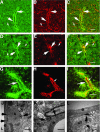
Figure 1
Figure 1
Localization of β8 in perivascular immature astrocytic cell processes in fetal brain and subcellular localization in adult rat brain hippocampus. A–C: Twenty-two week fetal cortex. D–I: Twenty-two week fetal periventricular zone. A, D, and G: Immunofluorescence localization of β8 surrounding blood vessels (large arrows) and in the surrounding developing cortex (small arrows). B: Nestin immunolocalization surrounding blood vessels (large arrows) and in the surrounding developing neuropil (small arrows). C: Co-localization of β8 (green) and nestin (red). Large arrows indicate co-localization (yellow) surrounding a blood vessel and small arrows indicate co-localization (yellow) in the developing neuropil. E: GFAP immunolocalization surrounding blood vessels (large arrows) and in the surrounding developing neuropil (small arrows). F: Co-localization of β8 (green) and GFAP (red). Large arrows indicate co-localization (yellow) surrounding a blood vessel and small arrows indicate co-localization (yellow) in the developing neuropil. H: CD34 immunolocalization in blood vessel endothelium (small arrows). I: Co-localization of β8 (green) and CD34 (red). The large arrow indicates β8 immunoreactivity in perivascular cell processes. The small arrow indicates CD34 immunofluorescence in the endothelium, which does not co-localize with β8. Shown are confocal images. J–L: Immunoelectron microscopy of adult rat brain showing β8 immunolocalization in glial processes. J: Horseradish peroxidase reaction product highlights an immunolabeled astrocytic process (A) adjacent to an endothelial cell (e) of a small blood vessel. Unlabeled terminals (uT) and an unlabeled dendritic spine (uS) are indicated. Arrows indicate patches of immunoperoxidase reaction product in the astrocytic process. K: Arrows indicate an immunolabeled astrocytic process in contact with the basement membrane of an endothelial cell (e), of a small blood vessel in the hippocampus of an adult rat. uT, unlabeled terminal. L: Arrow indicates an immunolabeled portion of an astrocytic process that is in close proximity to an endothelial cell (e) of a small blood vessel. uT, unlabeled terminal. Scale bars: 50 μm (C, F); 20 μm (I); 500 nm (J, K); 100 nm (L).
Figure 2
Figure 2
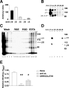
The integrin αvβ8 is expressed on the cell surface of human astrocytes, binds to and mediates cell adhesion to the LAP of TGF-β1. A: Flow cytometry of astrocytes stained with anti-integrin antibodies recognizing αvβ8, αvβ3, αvβ5, αvβ6, or α5 (n = 3). Shown is SE. Fluorescence intensity is shown in arbitrary units. B: Immunoprecipitation of 125I cell surface-labeled human astrocytes using anti-integrin subunit and complex specific antibodies against β1 (P5D2), αv (L230), β3 (AP3), αvβ5 (P1F6), αvβ6 (E7P6), or αvβ8 (14E5). Samples were resolved by 7.5% SDS-PAGE under nonreducing conditions. Shown are the migration of molecular weight markers on the right and the expected migration of integrin subunits on the left. C: LAP affinity chromatography of 125I surface-labeled human astrocyte lysates. Fractions were resolved by 7.5% SDS-PAGE under nonreducing conditions. Lanes 1 to 6, wash fractions; lanes 7 to 12, GRGESPK (1 mg/ml); lanes 13 to 16, GRGDSPK (1 mg/ml); lanes 17 to 20, ethylenediamine tetraacetic acid (10 mmol/L). Shown on the right are the molecular weight markers and the expected migration of integrin αv and β8 subunits. D: The RGD elution fractions were pooled and immunoprecipitated using anti-integrin subunit and complex specific antibodies against β1 (P5D2), αv (L230), β3 (AP3), αvβ5 (P1F6), αvβ6 (E7P6), or αvβ8 (14E5). Shown on the right are the molecular weight markers and the expected migration of integrin αv and β8 subunits. E: Astrocyte adhesion to immobilized LAP in the presence of no inhibitor (open bar), anti-αv (horizontal lines), or GRRGDLATIH (vertical lines). Shown is SE. *, P < 0.05; **, P < 0.001.
Figure 3
Figure 3
The activation and release of active TGF-β by astrocytes or freshly dissociated fetal brain cells is mediated by the integrin-αvβ8 and is metalloprotease-dependent. A: TGF-β bioassay of active TGF-β produced by astrocytes. Human astrocytes were co-cultured with TMLC TGF-β reporter cells, which stably express a portion of the plasminogen activator inhibitor-1 promoter driving the luciferase minigene, in the presence of no inhibitor (open bar), anti-β8 (horizontal lines), or anti-pan-TGF-β (filled bar). **, P < 0.001. B: TGF-β bioassay of active TGF-β released into the supernatants of human astrocytes. Supernatants from astrocytes in suspension were treated either with no inhibitor (open bar), anti-β8 (horizontal lines), anti-pan-TGF-β (filled bar), or GM6001, a pan-metalloprotease inhibitor (vertical lines) and cell-free supernatants were applied to TMLC cells. **, P < 0.001. C: Integrins known to interact with RGD that are expressed by freshly dissociated fetal brain cells as determined using flow cytometry using integrin heterodimer or subunit-specific antibodies to αvβ8, β3, αvβ5, αvβ6, or α5 (n = 3). Shown is mean fluorescence intensity. D: TGF-β bioassay of active TGF-β released into the supernatants of freshly dissociated fetal brain cells. Supernatants from freshly disaggregated fetal brain cells in suspension were treated either with no inhibitor (open bar), anti-β8 (horizontal lines), anti-pan-TGF-β (filled bar), or GM6001, a pan-metalloprotease inhibitor (vertical lines) and cell-free supernatants were applied to TMLC cells. Shown in A, B, and D are the fold increases of luciferase activity over the TMLC baseline activity. Shown is SE. *, P < 0.05; **, P < 0.001.
Figure 4
Figure 4
αvβ8-mediated activation of TGF-β by astrocytes inhibits endothelial migration in fibrin gels. A polyoma middle T-transformed murine brain endothelial cell line (bEND.3) was retrovirally transduced with a retrovirus encoding the enhanced green fluorescence protein (GFP) and was allowed to form a confluent monolayer on porcine collagen-coated microcarrier beads. Endothelial cell-coated beads alone (A, B, G, H, M, N, S, T), or endothelial cell-coated beads co-cultured with astrocyte-coated beads (C, D, I, J, O, P, U, V), or astrocyte-coated beads alone (E, F, K, L, Q, R, W, X) were embedded into fibrin gels containing either no additions (A–F), recombinant active TGF-β (G–L), anti-TGF-β1 (M–R), or anti-β8 (S–X). The assay was allowed to proceed 48 hours. The endothelial cells are phase and GFP-bright and the astrocytes are phase-bright and GFP-negative. Shown are representative phase (A, C, E, G, I, K, M, O, Q, S, U, W) and fluorescent (GFP) fields (B, D, F, H, J, L, N, P, R, T, V, X).
Figure 5
Figure 5
Gene profiling of endothelial cells reveals that key angiogenic genes are regulated by TGF-β. A: Real-time PCR of the murine endothelial bEND.3 cells using mouse-specific primers to thrombospondin-1, plasminogen activator inhibitor-1, urokinase plasminogen activator receptor, vascular endothelial growth factor-A, and angiopoietin-1 and -2, were used for real-time PCR to determine the relative abundance of angiogenic genes in untreated (open bars) or TGF-β1-treated (closed bars) cells. Shown are the relative gene copy numbers calculated using two species-specific housekeeping genes (chosen from five that showed the least variance between treatments) using geNorm. Shown is SE (n = 3).
Figure 6
Figure 6
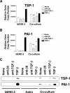
Astrocytic integrin αvβ8-mediated activation of TGF-β is sufficient to influence endothelial gene and protein expression of key anti-angiogenic genes. A and B: Real-time PCR of mouse bEND.3 cells alone or in co-culture with human astrocytes using mouse-specific primers to thrombospondin-1 (TSP-1) (A) or to plasminogen activator inhibitor-1 (PAI-1) (B). Cells were treated with nothing (open bars), anti-β8 (horizontal lines), anti-pan-TGF-β (vertical lines), or recombinant active TGF-β (filled bars). Shown are the relative gene copy numbers. Shown is SE (n = 3). *, P < 0.05; **, P < 0.001. C: Detection of thrombospondin-1 (TSP-1, top) or plasminogen activator inhibitor-1 (PAI-1, bottom) by Western blotting of supernatants of bEND.3, astrocytes, or co-cultures of bEND.3 and astrocytes treated with anti-β8, anti-pan-TGF-β, or recombinant active TGF-β. Shown is a representative experiment (n = 3).
Figure 7
Figure 7
Model of astrocytic integrin αvβ8-mediated activation of TGF-β as an angiogenic control switch. Integrin αvβ8 expressed in astrocyte end-feet binds to latent-TGF-β (L-TGF-β) localized in the basal lamina surrounding cerebral brain vessels, potentially stabilizing astrocyte end-feet association with blood vessels. Through a metalloproteolytic cleavage event, L-TGF-β bound to αvβ8 is activated and released allowing active TGF-β to diffuse to the abluminal surface of the endothelial cells where it can bind to TGF-β receptors and stimulate TGF-β signaling, leading to up-regulation of the anti-angiogenic genes, plasminogen activator inhibitor-1 (PAI-1) and thrombospondin-1 (TSP-1), which inhibit local fibrinolysis, cell migration, and proliferation. In this model, the homeostatic function of αvβ8-TGF-β interaction would be to stabilize the cerebral vasculature, and loss or gain of αvβ8 function could potentially lead to destabilization of cerebral vessels and angiogenesis or vascular wall thickening, respectively.

Similar articles

Cited by

References

    1. Ballabh P, Braun A, Nedergaard M. The blood-brain barrier: an overview: structure, regulation, and clinical implications. Neurobiol Dis. 2004;16:1–13. - PubMed
    1. Simard M, Arcuino G, Takano T, Liu QS, Nedergaard M. Signaling at the gliovascular interface. J Neurosci. 2003;23:9254–9262. - PMC - PubMed
    1. Pardridge WM. Blood-brain barrier biology and methodology. J Neurovirol. 1999;5:556–569. - PubMed
    1. Martin JS, Dickson MC, Cousins FM, Kulkarni AB, Karlsson S, Akhurst RJ. Analysis of homozygous TGF beta 1 null mouse embryos demonstrates defects in yolk sac vasculogenesis and hematopoiesis. Ann NY Acad Sci. 1995;752:300–308. - PubMed
    1. van den Driesche S, Mummery CL, Westermann CJ. Hereditary hemorrhagic telangiectasia: an update on transforming growth factor beta signaling in vasculogenesis and angiogenesis. Cardiovasc Res. 2003;58:20–31. - PubMed

Publication types

MeSH terms

LinkOut - more resources