. 2005 Jun;166(6):1895-905.
doi: 10.1016/S0002-9440(10)62498-4.

Prevention of neutrophil extravasation by hepatocyte growth factor leads to attenuations of tubular apoptosis and renal dysfunction in mouse ischemic kidneys

Affiliations

Prevention of neutrophil extravasation by hepatocyte growth factor leads to attenuations of tubular apoptosis and renal dysfunction in mouse ischemic kidneys

Shinya Mizuno et al. Am J Pathol. 2005 Jun.

Abstract

Ischemia and reperfusion (I/R) injuries occur in numerous organs under pathophysiological conditions. In this process, neutrophils play important roles in eliciting parenchymal injuries. Using a murine model of renal I/R, we show that hepatocyte growth factor (HGF) is a natural ligand that inhibits endothelial injuries and neutrophil extravasation. In mice after renal I/R, plasma HGF levels increased, along with c-Met/HGF receptor phosphorylation in the vascular endothelium. However, this c-Met activation was transient, associated with a decrease in endogenous HGF level, and followed by neutrophil infiltration and renal dysfunction. Suppression of endothelial c-Met phosphorylation by anti-HGF IgG led to rapid progressions of neutrophil extravasation, tubular apoptosis, and renal dysfunction. Inversely, enhancement of the c-Met activation by exogenous HGF blocked endothelial/tubular apoptotic injuries and acute renal failure. In this process, HGF prevented endothelial nuclear factor kappaB activation and inhibited induction of an adhesion molecule (ICAM-1), resulting in attenuated vascular edema and neutrophil infiltration. Thus, we conclude that 1) the HGF/c-Met system of endothelial cells confers an initial barrier to block neutrophil infiltration, and 2) transient and insufficient HGF production allows manifestation of postischemic renal failure. Our study provides a rationale for why HGF supplementation elicits therapeutic effects in ischemic kidneys.


Figures
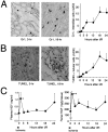
Figure 1
Figure 1
Changes in HGF levels during progression of neutrophil infiltration and tubular apoptosis in mice after renal I/R. A: Changes in the number of neutrophils (identified as Gr1-positive interstitial cells per ×200 field), noted in interstitial spaces of I/R-undergone kidneys (original magnification, ×330). The means of infiltrated neutrophils in each time point were calculated based on individual values (n = 6), which were determined on at least 20 fields per mouse. B: Alterations of tubular epithelial cell apoptosis (as determined by TUNEL staining) in ischemic kidneys (×330). C: Changes of plasma and renal HGF levels (as determined by a rodent HGF-specific ELISA system) after the release from warm ischemia. Statistical differences: *P < 0.05, **P < 0.01 versus pre-ischemia group (ie, 0-hour). For abbreviations, see text.
Figure 2
Figure 2
Acceleration of renal injuries and dysfunction in ischemic mouse kidneys by an anti-HGF IgG. A: Suppression of c-Met protein expression and phosphorylation by an antibody specific for rodent HGF. Left: Renal tissues were collected 1 hour after the I/R treatment, and then renal extract was immunoprecipitated with an anti-c-Met IgG and probed on PVDF membranes with an anti-phosphospecific c-Met IgG, as reported previously. Right: The band density was quantified using a densitometric analysis, with aid of computer software (NIH image). Data are expressed as mean ± SD (n = 4). **P < 0.01 versus normal IgG. B: Effects of the HGF-neutralizing treatment on BUN and plasma creatinine (Cr) levels. Mice were treated with anti-HGF IgG immediately after the surgical treatment of renal I/R and killed 8-hour postischemia. Data are shown as mean ± SD (n = 6). *P < 0.05, **P < 0.01 versus normal IgG group. C: Aggravating effects of anti-HGF IgG on tubular apoptosis in the renal I/R-undergone mice (8 hours postischemia). Representative findings of the apoptotic tubules in TUNEL-stained sections (×160). D: Acceleration of interstitial neutrophil infiltrations in the kidneys at 8 hours after HGF neutralization, as evidenced by counting Gr1-positive interstitial cells per ×200-field.
Figure 3
Figure 3
c-Met localization in mouse ischemic kidneys. A: Western blotting analysis for c-Met expression levels in kidneys after I/R. β-actin was detected as an internal control. Representative data were shown, and the density was quantified using a densitometric analysis, with aid of computer software (NIH image). B: Immunohistochemical findings of phosphorylated c-Met/HGF receptor in vascular endothelial cells in mice before and after renal I/R (original magnification: ×360). Renal tissues were fixed in 70% ethanol and then subjected to a process for paraffin embedding. Anti-phosphorylated c-Met IgG (pMet-1234/1235) was applied on the dewaxed sections, followed by an ABC immuno-peroxidase method. Insets: Normal rabbit IgG was applied on renal tissues (1 and 18 hours postischemia) as a negative control. Arrowheads: Positive signals in tubules.
Figure 4
Figure 4
Inhibitory effect of HGF on neutrophil adhesion and ICAM-1/nuclear NF-κB location in vitro. A: HGF-mediated suppressions of adhesions between HUVEC and peripheral blood-derived neutrophils. HUVECs were incubated for 3 hours with TNF-α (5 ng/ml). HGF (0–30 ng/ml) or vehicle (BSA) was pulsed into the HUVEC culture 30 minutes before adding TNF-α. After washing the cells with medium, neutrophils were transferred onto the HUVEC cell layer. The co-culture was kept for an additional 30 minutes, and then neutrophil adhesion was quantified using a MPO assay. B: Immunoblot analysis of ICAM-1 in whole-HUVEC cell lysates (top), which were harvested 3 hours after adding TNF-α (5 ng/ml), HGF (30 ng/ml) plus TNF-α, or HGF. The remaining HUVECs were fixed on a glass disk with 70% ethanol and then stained using an anti-ICAM-1 IgG (bottom). C: Inhibitory effects of HGF on TNF-α-induced NF-κB activation. Top: Immunoblot analysis of NF-κB (p65) in the nuclear extraction of HUVEC, incubated for 3 hours. Middle: Changes in nuclear location of NF-κB, as revealed by immunocytochemistry. The positive percentage of NF-κB-stained nucleoli were counted in >1000 cells per well (n = 6). Bottom: DNA-binding ELISA for detecting activated NF-κB in the whole-cell lysates of HUVECs. The culture condition was the same as in Figure 4A. Data were shown as means ± SD (n = 6). **P < 0.01 versus BSA; #P < 0.05 versus TNF-α alone.
Figure 5
Figure 5
Changes in plasma human HGF levels during administrations of rh-HGF to mice after renal ischemia. A: An experimental protocol of rh-HGF supplement therapy in the mouse model of postischemic acute renal failure. rh-HGF (or saline) was subcutaneously injected into mice immediately after renal I/R, and s.c. injections were repeated at an interval of 6 hours. The mice were killed at 3 and 18 hours to determine the effect of HGF on postischemic renal damages. B: Time course changes in plasma human HGF levels at 1, 3, and 18 hours after start of rh-HGF supplement therapy. The plasma was obtained from postorbital veins, and human HGF levels were determined using a human HGF-specific ELISA kit. S, saline; H, rh-HGF; n.d., not detectable.
Figure 6
Figure 6
HGF-induced suppressions of endothelial nuclear NF-κB and ICAM-1 expressions, related to attenuated vascular injuries in mice. A: Immunohistochemical detections of phosphorylated c-Met and nuclear NF-κB in renal vessels of the ischemic mice. The kidneys were collected at 3 hours after renal I/R and then subjected to immunohistochemistry for phosphorylated c-Met as well as for NF-κB (brown signals) and CD-31 (red signals), respectively. Arrowheads, vascular endothelium. #, tubular epithelial areas. Top inset: The time-matched renal tissue was immunostained with normal rabbit IgG and anti-CD31 mouse IgG (ie, negative control). Bottom inset: Expanded finding of the NF-κB-positive nuclear staining. B: Changes in endothelial ICAM-1 scores in kidneys, as determined by histological scores. Renal tissues were sampled before or 3 hours after renal I/R and then subjected to immunohistochemical procedures. Typical findings of endothelial ICAM-1 expression were shown. Arrowheads, endothelial surface. C: Suppression of endothelial apoptosis by rh-HGF in renal tissues, obtained at 3 hours after renal I/R. The kidneys were fixed in cold 70% ethanol, sectioned, and immunostained using an anti-mouse CD31 IgG. The CD31-stained tissue sections were refixed in 10% formalin and then subjected to TUNEL staining, as reported. D: Changes in peri-vascular edematous lesions (H&E staining). Arrowheads, edematous lesions. #, inflammatory cell accumulation. All data are shown as means ± SD (n = 6). In these experiments, statistical differences are expressed as *P < 0.05; **P < 0.01.
Figure 7
Figure 7
Preventive effects of rh-HGF supplement therapy toward neutrophil infiltration, tubular apoptosis, and renal dysfunction in mice that have undergone renal I/R. A: Changes in neutrophil accumulations in interstitial spaces of ischemic kidneys, treated with or without rh-HGF therapy (Gr1 staining, ×300). Renal tissues were collected at 3 and 18 hour after the renal I/R challenge and then subjected to immunohistochemical procedures. Furthermore, granulocyte-specific MPO activities were measured in a part of samples used in immunohistochemistry. In this assay, the saline control level (3 hours postischemia) was defined as 100%. B: Decrease in tubular apoptosis levels and of BUN levels in the mouse model of renal I/R injury by rh-HGF treatment. Data are shown as means ± SD (n = 6). *P < 0.05; **P < 0.01 compared with the saline-injected control group.

Similar articles

Cited by

References

    1. Beekhuizen H, van-de Gevel JS. Endothelial cell adhesion molecules in inflammation and postischemic reperfusion injury. Transplant Proc. 1998;30:4251–4256. - PubMed
    1. Jordan JE, Zhao ZQ, Vinten-Johansen J. The role of neutrophils in myocardial ischemia-reperfusion injury. Cardiovasc Res. 1999;43:860–878. - PubMed
    1. Martin TR. Neutrophils and lung injury: getting it right. J Clin Invest. 2002;110:1603–1605. - PMC - PubMed
    1. Lauriat S, Linas SL. The role of neutrophils in acute renal failure. Semin Nephrol. 1998;18:498–504. - PubMed
    1. Scarabelli T, Stephanou A, Rayment N, Pasini E, Comini L, Curello S, Ferrari R, Latchman D. Apoptosis of endothelial cells precedes myocyte cell apoptosis in ischemia and reperfusion injury. Circulation. 2001;104:253–256. - PubMed

Publication types

MeSH terms

Substances