. 2005 Jun 15;24(12):2114-26.
doi: 10.1038/sj.emboj.7600696. Epub 2005 May 26.

v-SNAREs control exocytosis of vesicles from priming to fusion

Affiliations

v-SNAREs control exocytosis of vesicles from priming to fusion

Maria Borisovska et al. EMBO J. .

Abstract

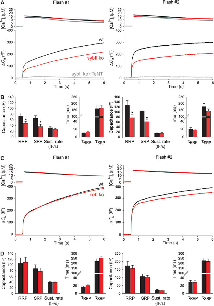
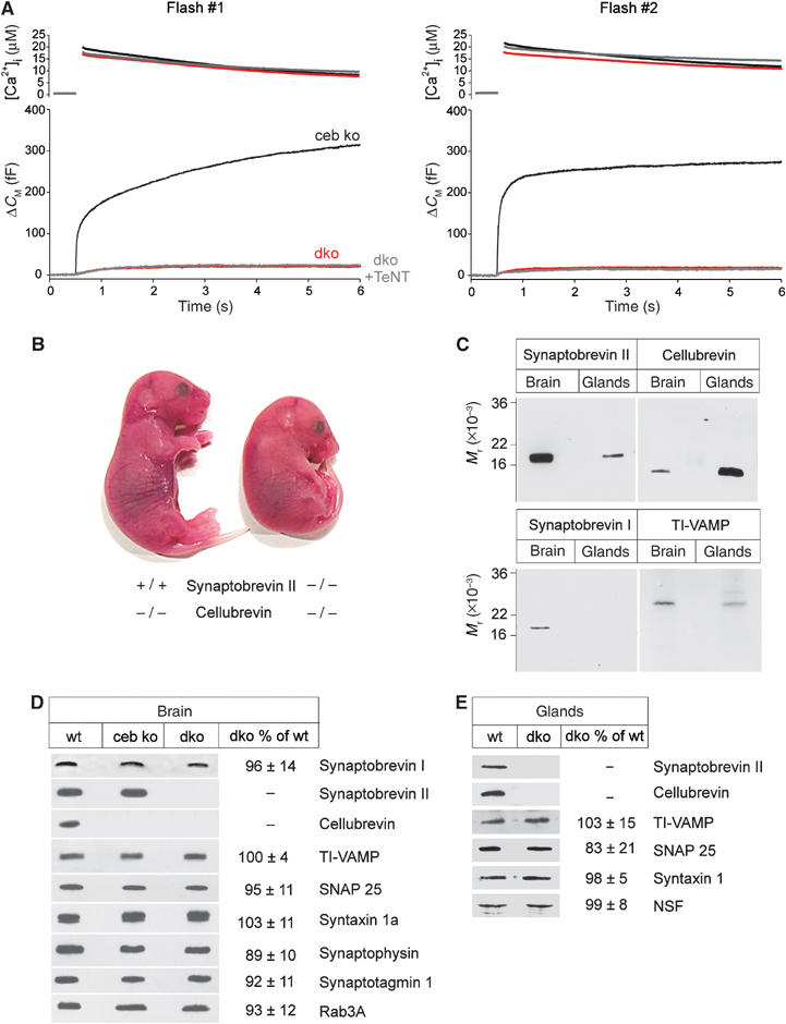
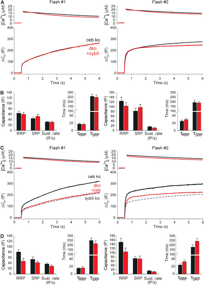
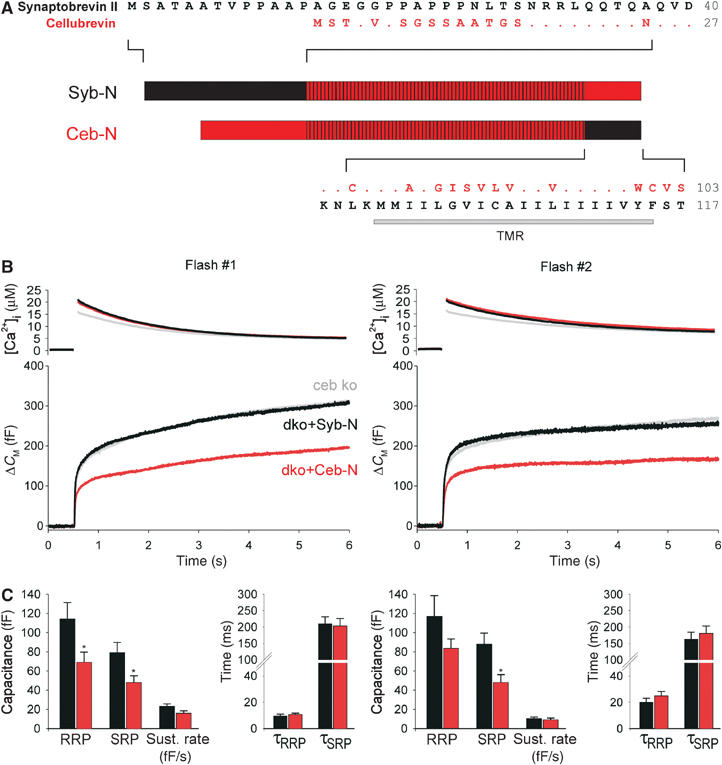
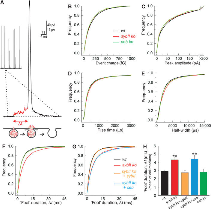
SNARE proteins (soluble NSF-attachment protein receptors) are thought to be central components of the exocytotic mechanism in neurosecretory cells, but their precise function remained unclear. Here, we show that each of the vesicle-associated SNARE proteins (v-SNARE) of a chromaffin granule, synaptobrevin II or cellubrevin, is sufficient to support Ca(2+)-dependent exocytosis and to establish a pool of primed, readily releasable vesicles. In the absence of both proteins, secretion is abolished, without affecting biogenesis or docking of granules indicating that v-SNAREs are absolutely required for granule exocytosis. We find that synaptobrevin II and cellubrevin differentially control the pool of readily releasable vesicles and show that the v-SNARE's amino terminus regulates the vesicle's primed state. We demonstrate that dynamics of fusion pore dilation are regulated by v-SNAREs, indicating their action throughout exocytosis from priming to fusion of vesicles.


Figures

Figure 1
Figure 1
Lack of sybII or ceb differentially affects secretion from chromaffin cells. (A) Averaged flash-induced [Ca2+]i levels (upper panel; flash at t=0.5 s) and corresponding capacitance responses (lower panel) of wild-type (wt; n=22), sybII ko (n=22) and sybII ko cells treated with tetanus toxin light chain (sybII ko+TeNT, n=6). Secretion is diminished in mutant cells but almost completely blocked upon additional poisoning with TeNT. Similar effects are observed for the second flash response given with a 2 min interval (right panels). Preflash [Ca2+]i levels (in μM): first flash: wt 0.31±0.02, sybII ko 0.32±0.01, sybII ko+TeNT 0.38±0.04; second flash: wt 0.43±0.04, sybII ko 0.38±0.02, sybII ko+TeNT 0.49±0.1. (B) Amplitudes of the two exocytotic burst components (RRP, SRP) and the rate of sustained release (fF/s) for control (black bars) and sybII ko cells (red bars). Compared with controls, the average RRP and SRP response of mutant cells is similarly reduced to 65 and 55% for the first flash and to 61 and 58% for the second flash, respectively. No change is observed for the time constants of the individual exocytotic burst components (τRRP, τSRP). *P<0.05 (Student's t-test). (C) Secretion of ceb ko cells is indistinguishable from that of wt cells. Preflash [Ca2+]i was the same for wt and ceb ko cells (in μM): first flash, 0.36±0.01; second flash, 0.45±0.02. (D) Neither the magnitude nor the kinetic properties are significantly changed in ceb ko cells (red bars) compared with controls (black bars).
Figure 2
Figure 2
Double v-SNARE deficiency abolishes Ca2+-triggered secretion from chromaffin cells. (A) The flash-evoked capacitance increase is almost abolished in dko cells (n=20). ceb ko cells (n=20) serve as littermate control in these experiments. The residual capacitance response of dko cells is resistant to TeNT poisoning (dko+TeNT, n=12). Preflash [Ca2+]i levels (in μM): first flash: ceb ko 0.34±0.01, dko 0.33±0.01, dko+TeNT 0.39±0.02; second flash: ceb ko 0.43±0.02, dko 0.41±0.02, dko+TeNT 0.54±0.04. (B) Appearance of double v-SNARE-deficient mutant mice at stage E18.5 compared with littermates (ceb ko). (C) Survey on the expression of v-SNARE proteins in embryonic adrenal glands and the brain of wild-type (wt) animals. Each lane is an immunoblot of total homogenate (sybII and ceb, 5 μg/lane; synaptobrevin I, 15 μg/lane; TI-VAMP, 25 μg/lane). Note that ceb, detectable in brain homogenate, likely derives from glia cells (Chilcote et al, 1995). (D) Immunoblot analysis of total brain homogenate from wt and dko mice illustrates no major changes in the expression level of other synaptic proteins. Homogenates were probed with antibodies raised against the indicated proteins. Signals were visualized with enhanced chemiluminescence. (E) Immunoblot of embryonic adrenal glands from wt and dko animals indicates no change in the expression pattern of other synaptic proteins in the absence of sybII and ceb. Quantification of the immunosignals from dko in panels D and E is given as mean percentage±s.d. compared with wt. Data were collected from three independent preparations of brain and gland homogenate.
Figure 3
Figure 3
Electron microscopy of chromaffin granules lacking sybII and ceb. (A) Exemplary electron micrographs of isolated mouse chromaffin cells from wild-type (wt) and dko animals. Scale, 2 μm. (B) dko cells exhibit the same density of chromaffin granules as found in ceb ko or wt cells. wt cells exhibit on average 156±13 granules/section. Data were collected from 16 wt, 17 ceb ko and 14 dko cells. (C) Size distribution of wt (black line, n=916), ceb ko (gray, n=1122) and dko granules (red, n=992). A Gaussian fitted to each of the distributions (not shown) was used to determine the parameters indicated in the text (mean±s.d.). (D) Subcellular distribution (bin width 50 nm) of chromaffin granules in wt (black), ceb ko (gray) and dko cells (red) measured as granule membrane to plasma membrane distance. Note that in dko cells, there is a slight (but nonsignificant) increase in the number of docked vesicles that may result from the observed blockade in exocytosis causing an accumulation of vesicles in the docked state. Inset: Morphologically docked granule in close proximity to the plasma membrane; intermembrane distance ∼10 nm. Scale, 100 nm.
Figure 4
Figure 4
v-SNARE dependence of depolarization-induced capacitance increase and catecholamine secretion. (A–C) Average CM responses from sybII ko (n=22), ceb ko (n=26) and dko cells (n=21) compared with the littermate control responses. Cells were stimulated with a train of 100 ms depolarizations (2.5 Hz, voltage protocol, upper panel). Note that wild-type (wt) cells show a stronger increase of the capacitance response than syb ko cells during the stimulus train. This is expected for a reduced SRP component in syb ko cells (as observed in the flash experiments), which occurs less synchronous with such a stimulus. Data were collected from more than 20 cells for each control group. (D) Mean amplitude of the Ca2+ current (ICa2+) and the corresponding CM increase (CM) for the first depolarization. CM was determined at the end of the capacitance recording interval. Data were normalized to the response of controls. The total capacitance increase recorded from dko cells at the end of the stimulus train amounts for less than 10% of the control response. (E–H) Representative amperometric recordings of catecholamine release induced by application of high K+-containing Ringer's solution (80 mM KCl) recorded for the indicated genotypes. Note that the response is reduced in sybII ko cells compared with wt cells and is abolished in dko cells. (I) Plot of the mean flash-evoked capacitance response (measured 5.5 s after stimulation) versus the mean number of amperometric events recorded for the different v-SNARE deficiencies. Data were normalized to the response of the corresponding control group. A linear regression (solid line) approximates the data (r2=0.98).
Figure 5
Figure 5
Expression of either v-SNARE, sybII or ceb, ‘rescues' secretion in dko cells. (A) Mean flash-evoked capacitance response of dko cells expressing sybII (dko+sybII, n=28) compared with controls (ceb ko, n=30). Preflash [Ca2+]i levels (in μM): first flash: ceb ko 0.40±0.01, dko+sybII 0.41±0.01; second flash: ceb ko 0.51±0.02, dko+sybII 0.61±0.03. Notably, secretion from dko cells is rescued within 4–6 h after start of virus-driven protein expression. This may be accomplished either by direct sorting of new v-SNAREs to pre-existing granules or by rapid de novo synthesis of granules. The latter scenario requires preferential recruitment of newly formed vesicles for exocytosis as observed by Duncan et al (2003). (B) Expression of sybII in dko cells (red bars) restores magnitude and kinetics of RRP and SRP as well as the sustained rate of secretion, control (black bars). (C) Average flash-evoked capacitance response of dko cells expressing ceb (dko+ceb, n=19) compared with controls (ceb ko, n=20). Preflash [Ca2+]i levels (in μM): first flash: ceb ko 0.35±0.01, dko+ceb 0.34±0.01; second flash: ceb ko 0.44±0.02, dko+ceb 0.47±0.02. The dashed line represents sybII ko measurement (from Figure 1A) indicating near identity with the dko+ceb phenotype. Note that the slight, but nonsignificant difference in the exocytotic burst size for the second flash response of dko+ceb and sybII ko cells is likely due to higher preflash [Ca2+]i levels in virus-transfected cells (0.47 μM) compared with nontransfected sybII ko cells (0.38 μM). (D) Expression of ceb in dko cells (red bars) partially restores secretion compared with controls (black bars). RRP and SRP sizes are ‘rescued' to 58 and 70% for the first flash and to 77 and 98% for the second flash, respectively. *P<0.05 compared with controls (Student's t-test).
Figure 6
Figure 6
The amino terminus of the v-SNARE protein determines the vesicle's priming stability. (A) Schematic presentation of sybII/ceb chimeras: Syb-N (aa 1–37 sybII+aa 25–103 ceb); Ceb-N (aa 1–24 ceb+aa 38–117 sybII). The hatched area illustrates the conserved region of the proteins. Dots indicate amino acids of ceb (red) that are identical to those of sybII (black). TMR, transmembrane region. (B) Mean flash-evoked capacitance response of ceb ko cells (gray, n=37), dko cells expressing the chimera syb-N (black, n=24) and those expressing the chimera ceb-N (red, n=24). Preflash [Ca2+]i levels (in μM): first flash: ceb ko 0.4±0.1, syb-N 0.44±0.02, ceb-N 0.42±0.02; second flash: ceb ko 0.54±0.03, syb-N 0.58±0.03, ceb-N 0.56±0.05. (C) Syb-N expression in dko cells (black bars) supports a larger pool of exocytosis-competent vesicles than Ceb-N expression (red bars). *P<0.05 (Student's t-test).
Figure 7
Figure 7
Lack of sybII prolongs the expansion time of the exocytotic fusion pore. (A) Exemplary amperometric event recorded with a carbon fiber in direct contact with the plasma membrane of a chromaffin cell. A ‘foot' signal (red line) precedes the main amperometric spike indicative for restricted transmitter efflux through a narrow fusion pore that slowly expands (t=‘foot' duration, see scheme) before bulk release, coinciding with more rapid pore dilation. (B–E) Characteristics of single spikes recorded from wild-type (wt; black), sybII ko (red) and ceb ko cells (green) displayed as cumulative frequency distributions for the indicated parameters. The properties of the amperometric signals from the v-SNARE-deficient cells are very similar compared with controls and curves partially overlap (values are given in Table I). (F) The ‘foot' duration of amperometric spikes is significantly longer in sybII ko (red) than in wt (black) or ceb ko cells (green). No change in the frequency of ‘foot' signals is observed (see Table I). (G) Expression of sybII (orange), but not of ceb (blue), in sybII ko cells (red) ‘rescues' the ‘foot' duration to wt levels (black). (H) Mean value of cell medians of ‘foot' duration for all studied conditions. *P<0.05 compared with wt cells.

References

    1. Albillos A, Dernick G, Horstmann H, Almers W, Alvarez de Toledo G, Lindau M (1997) The exocytotic event in chromaffin cells revealed by patch amperometry. Nature 389: 509–512 - PubMed
    1. Ashery U, Betz A, Xu T, Brose N, Rettig J (1999) An efficient method for infection of adrenal chromaffin cells using the Semliki Forest virus gene expression system. Eur J Cell Biol 78: 525–532 - PubMed
    1. Baumert M, Maycox PR, Navone F, De Camilli P, Jahn R (1989) Synaptobrevin: an integral membrane protein of 18,000 daltons present in small synaptic vesicles of rat brain. EMBO J 8: 379–384 - PMC - PubMed
    1. Bezzi P, Gundersen V, Galbete JL, Seifer G, Steinhauser C, Pilati E, Volterra A (2004) Astrocytes contain a vesicular compartment that is competent for regulated exocytosis of glutamate. Nat Neurosci 7: 613–620 - PubMed
    1. Bhattacharya S, Stewart BA, Niemeyer BA, Burgess RW, McCabe BD, Lin P, Boulianne G, O'Kane CJ, Schwarz TL (2002) Members of the synaptobrevin/vesicle-associated membrane protein (VAMP) family in Drosophila are functionally interchangeable in vivo for neurotransmitter release and cell viability. Proc Natl Acad Sci USA 99: 13867–13872 - PMC - PubMed

Publication types

MeSH terms