. 2005 Jun 15;24(12):2194-204.
doi: 10.1038/sj.emboj.7600683. Epub 2005 May 26.

Concerted mechanism of Swe1/Wee1 regulation by multiple kinases in budding yeast

Affiliations

Concerted mechanism of Swe1/Wee1 regulation by multiple kinases in budding yeast

Satoshi Asano et al. EMBO J. .

Abstract

In eukaryotes, entry into mitosis is induced by cyclin B-bound Cdk1, which is held in check by the protein kinase, Wee1. In budding yeast, Swe1 (Wee1 ortholog) is targeted to the bud neck through Hsl1 (Nim1-related kinase) and its adaptor Hsl7, and is hyperphosphorylated prior to ubiquitin-mediated degradation. Here, we show that Hsl1 and Hsl7 are required for proper localization of Cdc5 (Polo-like kinase homolog) to the bud neck and Cdc5-dependent Swe1 phosphorylation. Mitotic cyclin (Clb2)-bound Cdc28 (Cdk1 homolog) directly phosphorylated Swe1 and this modification served as a priming step to promote subsequent Cdc5-dependent Swe1 hyperphosphorylation and degradation. Clb2-Cdc28 also facilitated Cdc5 localization to the bud neck through the enhanced interaction between the Clb2-Cdc28-phosphorylated Swe1 and the polo-box domain of Cdc5. We propose that the concerted action of Cdc28/Cdk1 and Cdc5/Polo on their common substrates is an evolutionarily conserved mechanism that is crucial for effectively triggering mitotic entry and other critical mitotic events.


Figures

Figure 1
Figure 1
Hsl1 and Hsl7 are critical for Cdc5-dependent Swe1 phosphorylation and degradation. (A) Strain JC66 bearing YCplac22, pKL882 (GAL1-EGFP-CDC5), or pKL883 (GAL1-EGFP-cdc5(FAA)) was cultured in YEP-raffinose medium and arrested in S phase with hydroxyurea for 2.5 h before transferring to YEP-galactose supplemented with hydroxyurea for 3 h. Total cellular proteins were separated by 10% SDS–PAGE and then subjected to immunoblot analyses with anti-Swe1 and anti-GFP (for EGFP-Cdc5) antibodies. The Asterisk indicates a nonspecific protein crossreacting with the anti-Swe1 antibody. (B) Strains KLY1546 (wild type), KLY2868 (hsl1Δ), and KLY2872 (hsl7Δ) were arrested in S phase with hydroxyurea (S), or in M phase with nocodazole (M). Total cellular proteins were separated by 10% SDS–PAGE for subsequent immunoblotting analyses. (C–E) Strains KLY1546 (C), KLY2868 (D), or KLY2872 (E) were transformed with either YCplac22 (vector) or pKL882 (pGAL1-EGFP-CDC5). The resulting transformants were treated with hydroxyurea for 2.5 h and then transferred to YEP-galactose containing hydroxyurea (t=0). Total cellular proteins were separated in 8% low-bis SDS–PAGE. (F) Strains KLY1546 (wild type), KLY2868 (hsl1Δ), and KLY2872 (hsl7Δ) were transformed with either pSK754 (EGFP-CDC5) or pKL880 (GAL1-EGFP-CDC5ΔN). The resulting transformants were cultured overnight at 18°C (EGFP-Cdc5 signals were more stable at a low temperature) in YEP-glucose (for pSK754) or YEP-raffinose (for pKL880). Cells were treated with nocodazole at 18°C for 5 h (for pSK754) or for 4 h followed by 1 h induction in YEP-galactose containing nocodazole (for pKL880) before examination under a fluorescent microscope. Error bars indicate standard deviation.
Figure 2
Figure 2
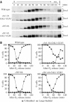
Mitotic cyclin-associated Cdc28 activity is required for proper Swe1 phosphorylation and degradation. (A) Strains 15D (isogenic wild type), SBY458 (cln1-3Δ GAL1-CLN3), SBY145 (clb5-6Δ), and SBY175 (clb1-4Δ GAL1-CLB1) were cultured in YEP-galactose at 30°C, arrested in G1 by α-factor treatment and then released into YEP-glucose containing nocodazole (see Materials and methods for details). Total cellular proteins prepared at each time point after α-factor release were separated by 8% low-bis SDS–PAGE. Asterisks indicate a crossreacting protein with anti-Swe1 antibody. (B) Budding indices were determined to monitor the cell cycle progression from the same samples as in panel A.
Figure 3
Figure 3
Clb2-Cdc28 and Cdc5 phosphorylate Swe1 synergistically in vitro. (A) Cyclin-associated Cdc28 complexes were reacted with GST-swe1(K473A) and histone H1 (HH1) as in vitro substrates in each reaction. The reaction mixtures were separated by 8% low-bis SDS–PAGE (upper gel) to detect Swe1 mobility shift and 10% normal SDS–PAGE (lower gel) to retain HH1 in the gel, and then subjected to autoradiography. WT, GST-Cdc28; DN, GST-cdc28(D145N). (B, C) GST-Cdc28/His6-Cks1/MBP-Clb2, T7-HA-Cdc5-Flag, and T7-HA-Cla4-Flag were reacted with kinase-inactive GST-swe1(K473A) in the presence of 50 μM ATP (10 μCi of [γ-32P]ATP) at 30°C. Reactions were terminated at the indicated time points and then separated by 8% low-bis SDS–PAGE. After staining with Coomassie, the gel was subjected to autoradiography. Cdc5 WT, T7-HA-Cdc5-FLAG; cdc5NA, kinase-inactive T7-HA-cdc5(N209A)-FLAG; Cla4 WT, T7-HA-Cla4-FLAG; cla4KA, kinase-inactive T7-HA-cla4(K594A)-FLAG. (D, E) The bands corresponding to GST-swe1(K473A) from the reactions were excised and the incorporated 32P was quantified. Error bars indicate standard deviation.
Figure 4
Figure 4
Priming of Swe1 by Clb2-Cdc28 enhances Cdc5-dependent Swe1 phosphorylation in vitro. (A) Scheme of the two-step kinase reactions. (B, C) Bead-bound GST-swe1(K473A) was primed by the indicated enzymes in the first reaction. After washing out the remaining ATP, second enzymes were added as indicated to carry out labeling reactions (see Materials and methods for details). Samples were separated by 8% low-bis SDS–PAGE and then analyzed. The asterisk indicates a contaminating protein copurified with Cdc5 and Cla4 from Sf9 cells. Cdc5, T7-HA-Cdc5-FLAG; cdc5(N209A), T7-HA-cdc5(N209A)-FLAG; Cdc28, MBP-Clb2/GST-Cdc28/His6-Cks1 complex; cdc28(D145N), MBP-Clb2/GST-cdc28(D145N)/His6-Cks1 complex; Cla4, T7-HA-Cla4-FLAG; cla4(K594A), T7-HA-cla4(K594A)-FLAG. (D, E) Cdc5 activation assays with Clb2-Cdc28. Reactions were carried out as described in panel D and analyzed as above. Cdc28 W, MBP-Clb2/GST-Cdc28/His6-Cks1 complex; Cdc28 D, MBP-Clb2/GST-cdc28(D145N)/His6-Cks1 complex; Cdc5 W, T7-HA-Cdc5-FLAG; Cdc5 N, T7-HA-cdc5(N209A)-FLAG.
Figure 5
Figure 5
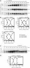
Requirement of mitotic Clb-Cdc28 activity for Cdc5-dependent Swe1 phosphorylation and degradation in vivo. Strain KLY5426 (cdc28-as1 cdc5Δ+ pGAL1-cdc5-1) transformed with either pKL321 (YCplac22-CDC5) or YCplac22 (control vector) was cultured in YEP-galactose, arrested in G1, and then released into YEP-glucose medium containing nocodazole (see Materials and methods for details). Either control DMSO or 0.5 μM of 1NM-PP1 was added to the cultures as indicated. (A, C) Total cellular proteins were separated by 8% low-bis SDS–PAGE. Asterisks indicate a nonspecific protein crossreacting with anti-Swe1 antibody. (B, D) To monitor cell cycle progression, samples harvested in panel A or C were counted for budding indices.
Figure 6
Figure 6
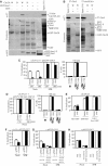
Clb2-Cdc28 is critical for Cdc5-dependent Swe1 regulation. (A, B) GST-swe1(K473A) primed by either GST-Cdc28/His6-Cks1/MBP-Clb2, catalytically inactive GST-cdc28(D145N)/His6-Cks1/MBP-Clb2, or control buffer was incubated with clarified cellular lysates from either Sf9 cells expressing T7-HA-cdc5(N209A)-FLAG (A) or bacterial cells expressing T7-cdc5(N209A)-His6 or T7-cdc5(FAA)-His6 (B) (see Materials and methods for details). After pull-down, proteins were separated by 10% normal SDS–PAGE to retain the GST ligand in the gel (as a result, the migration pattern of phosphorylated GST-Swe1 appeared slightly different from that in 8% low-bis SDS–PAGE). The migration difference of the GST ligand in panel A does not reflect its actual mobility difference in the gel. W, GST-Cdc28/His6-Cks1/MBP-Clb2; D, GST-cdc28(D145N)/His6-Cks1/MBP-Clb2; GST-Swe1-P, phosphorylated GST-swe1(K473A) (arrows). As input, 1% of the clarified cellular lysates was used. (C, D) Strain KLY5401 transformed with either centromeric pSK754 (pEGFP-CDC5) (C) or centromeric pKL2438 (pYFP-CDC5ΔC-CDC12) (D) was cultured overnight, arrested by α-factor treatment, and then released into YEP-glucose containing nocodazole. Either DMSO or 0.5 μM of 1NM-PP1 was added into the culture 20 min after release. Samples harvested at the indicated time points after release were fixed and then examined. Greater than 500 cells were counted from duplicated experiments. (C, D, right panels) DAPI morphology of the 180 min samples in panels C and D was quantified. Treatment of cells with 1NM-PP1 induced elongated bud morphologies. Error bars indicate standard deviation. (E) Strain KLY5401 (cdc28-as1) bearing either centromeric YCpT-HSL1-GFP, YCpT-GFP-HSL7, or YCpLG-SWE1-GFP (GAL1-promoter controlled) was cultured in YEP-glucose (for Hsl1-GFP and GFP-Hsl7) or YEP-raffinose (for GAL1-Swe1-GFP) at 23°C and then arrested with nocodazole for 3 h. Cells were then treated with 0.5 μM of 1NM-PP1 for 2 h either in the same medium (for Hsl1-GFP and GFP-Hsl7) or after transferring to YEP-galactose containing nocodazole (for GAL1-Swe1-GFP). Samples were examined as in panels C and D. A low efficiency of Hsl1-GFP localization in comparison to GFP-Hsl7 localization is likely due to weaker Hsl1-GFP fluorescent signals. Error bars indicate standard deviation. (F) Strain KLY4362 (swe1Δ) transformed with either a centromeric pSWE1 or control vector was additionally transformed with pSK754 (pEGFP-CDC5). The resulting transformants were cultured at 23°C and arrested with hydroxyurea for 6 h (the condition where Swe1 is abundant and a significant level of EGFP-Cdc5 signal is detectable at the bud neck) before fixation. Error bars indicate standard deviation. (G, H) Localization of EGFP-Cdc5 in the indicated genetic background was examined as in panel F. Y19F, CDC28 Y19F allele.
Figure 7
Figure 7
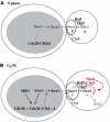
Model illustrating the multi-kinase-dependent Swe1 phosphorylation and degradation during the cell cycle. (A) In S phase, Cla4 is primarily responsible for Swe1 phosphorylation. Swe1 is less abundant at this stage, and thus is shown in plain letters. Swe1-dependent Cdc28 Tyr19 phosphorylation occurs as early as S phase. (B) In G2, Swe1 becomes abundant (Swe1 in bold letters) and more potently inhibits Clb-Cdc28. Prior to mitotic entry, a rise in Clb-Cdc28 activity as the level of mitotic Clb increases tips the balance to trigger Clb-Cdc28-dependent Swe1 phosphorylation. This step is critical both for promoting Swe1–Cdc5 interaction and for the subsequent Cdc5-dependent Swe1 phosphorylation at the bud neck (see text for details). Dotted lines indicate interactions among the proteins. Triple dots between Hsl1–Hsl7 and neck–Cdc5 indicate a putative interaction between these proteins (see online version for color figure).

References

    1. Bishop AC, Ubersax JA, Petsch DT, Matheos DP, Gray NS, Blethrow J, Shimizu E, Tsien JZ, Schultz PG, Rose MD, Wood JL, Morgan DO, Shokat KM (2000) A chemical switch for inhibitor-sensitive alleles of any protein kinase. Nature 407: 395–401 - PubMed
    1. Booher RN, Deshaies RJ, Kirschner MW (1993) Properties of the Saccharomyces cerevisiae wee1 and its differential regulation of p34cdc28 in response to G1 and G2 cyclins. EMBO J 12: 3417–3426 - PMC - PubMed
    1. Elia AE, Cantley LC, Yaffe MB (2003) Proteomic screen finds pSer/pThr-binding domain localizing Plk1 to mitotic substrates. Science 299: 1228–1231 - PubMed
    1. Hwang WW, Silver PA (2001) The Saccharomyces cerevisiae cyclin Clb2p is targeted to multiple subcellular locations by cis- and trans-acting determinants. J Cell Sci 114: 589–597 - PubMed
    1. Kaiser P, Sia RAL, Bardes EGS, Lew DJ, Reed SI (1998) Cdc34 and the F-box protein Met30 are required for degradation of the Cdk-inhibitory kinase Swe1. Genes Dev 12: 2587–2597 - PMC - PubMed

Publication types

MeSH terms