. 2005 Jun 15;24(12):2096-103.
doi: 10.1038/sj.emboj.7600675. Epub 2005 May 26.

Bax forms multispanning monomers that oligomerize to permeabilize membranes during apoptosis

Affiliations

Bax forms multispanning monomers that oligomerize to permeabilize membranes during apoptosis

Matthew G Annis et al. EMBO J. .

Abstract

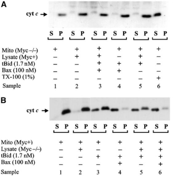
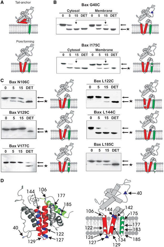
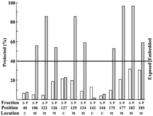
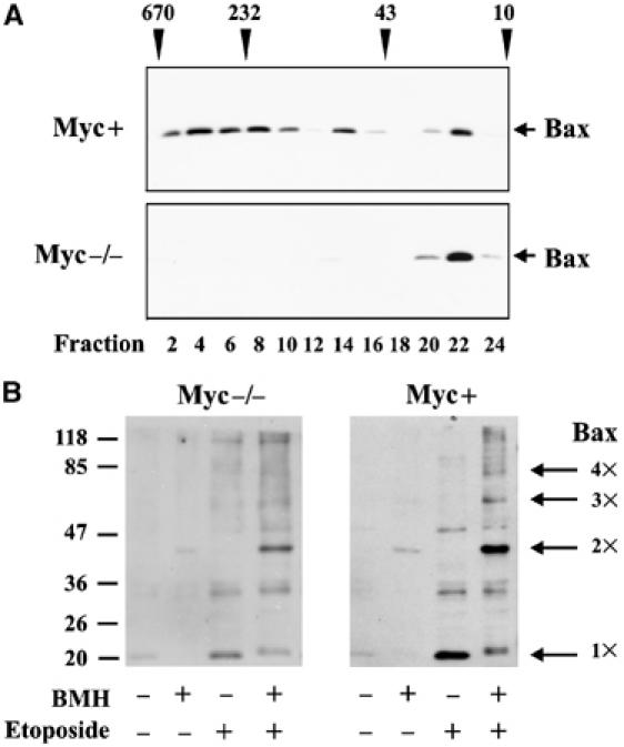
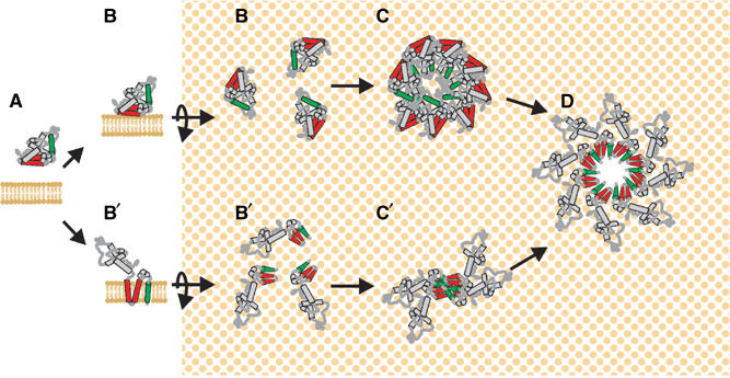
Bax promotes cell death by permeabilizing mitochondrial outer membranes by an unresolved mechanism. However, in cells lacking the gene c-myc, membrane permeabilization by Bax is blocked by changes in the mitochondria that prevent Bax oligomerization. Drug-treated c-myc null cells and cells expressing Myc were used to map the topology of Bax in membranes prior to and after mitochondrial permeabilization. Chemical labeling of single cysteine mutants of Bax using a membrane bilayer impermeant cysteine-specific modifying agent revealed that Bax inserted both the 'pore domain' (helices alpha5-alpha6), and the tail-anchor (helix alpha9) into membranes prior to oligomerization and membrane permeabilization. Additional topology changes for Bax were not required in Myc-expressing cells to promote oligomerization and cytochrome c release. Our results suggest that unlike most pore-forming proteins, Bax membrane permeabilization results from oligomerization of transmembrane monomers rather than concerted insertion of the pore domains of a preformed oligomer.


Figures

Figure 1
Figure 1
Mitochondria from Myc−/− cells are more resistant than mitochondria from Myc+ cells to induction of cytochrome c release by 100 nM Bax and 1.7 nM tBid. Mitochondria isolated from (A) Myc−/− or (B) Myc+ cells were incubated with tBid, Bax and cytosol as indicated. Cytochrome c release was assayed by pelleting the mitochondria and visualizing the cytochrome c in the supernatant (S) and pellet (P) fractions by SDS–PAGE and immunoblotting. As a control, cytochrome c was released from mitochondria by solubilization using the nonionic detergent Triton X-100 (TX-100).
Figure 2
Figure 2
In Myc−/− cells, both the pore-forming and tail-anchor domains of membrane-bound Bax are embedded in the bilayer. (A) Membrane-bound Bax is predicted to adopt either a tail-anchor or pore-forming topology. Helices are diagrammed as solid rods, the tail-anchor (helix 9) is green and the ‘pore-forming' helices (5–6) are red. (B, C) Cytosolic and membrane fractions isolated from etoposide-treated Myc−/− cells expressing the indicated mutants were labeled with IASD (time indicated above the lanes) and labeling was assessed by isoelectric focusing and immunoblotting. Arrows indicate the migration positions of Bax and * indicates IASD-labeled Bax. The location of the single cysteine is indicated as a triangle on the diagram at the right of each panel. Overhead arrows indicate bands representing residues protected from IASD. (B) Labeling of cytoplasmic (Bax G40C) and integrated (Bax I175C) controls. In cytosol and membrane fractions, Bax G40C was labeled by IASD. The cytosolic form of Bax I175C was labeled by IASD, but in membrane fractions this residue was protected from labeling until the bilayer was solubilized with detergent (DET). (C) Labeling reactions for membrane fractions from Myc−/− cells expressing the indicated single cysteine Bax mutants. Interpretive diagrams are presented to the right of each panel. (D) Comparison of the solution structure and the membrane topology of Bax in etoposide-treated Myc−/− and Myc+ cells. Blue residues on the solution structure (left) indicate positions of single cysteine mutants. In the topology diagram (right), closed triangles represent residues modified similarly by IASD in cytosolic and membrane-bound Bax. Open triangles represent residues that were protected from IASD by the membrane.
Figure 3
Figure 3
Quantification of labeling efficiency of Bax single cysteine mutants. Open and gray bars indicate supernatant (S) and membrane pellet (P) fractions from etoposide-treated cells, respectively. The position of the cysteine residue is indicated below the lanes. The deduced location of the residue is indicated as exposed to cytoplasm (C) or inserted into the membrane (M). The line at 40% protected represents an arbitrary cutoff that can be used to distinguish exposed cytoplasmic from membrane-embedded cysteines without reference to the supernatant fractions. The approximate error (standard deviation) for each measurement is 10%.
Figure 4
Figure 4
Myc expression is required for Bax oligomerization. Oligomerization of Bax was measured by (A) gel filtration chromatography and (B) crosslinking with BMH of membrane fractions. The membrane fraction was solubilized in CHAPS for chromatography. Fraction number and the migration positions of molecular weight standards are indicated below and above the immunoblots in (A). Crosslinking was performed in cell lysates prepared by nitrogen cavitation and then membrane fractions were collected by centrifugation. Migration positions for molecular weight standards are shown to the left of the panels. Deduced migration positions for Bax monomer and oligomers are indicated to the right of the panels in (B).
Figure 5
Figure 5
Comparison of pore-forming models for Bax. (A) In untreated cells, Bax is a cytoplasmic monomer. In the model based on cytolytic toxins, (B) Bax binds to membranes peripherally or by insertion of helix 9. (C) Membrane-bound monomers oligomerize to generate a structure capable of forming a pore in the membrane. (D) A concerted conformational change inserts helices 5–6 and 9 (unless helix 9 is already embedded) and reorganizes lipids to form a large hole in the membrane. In the oligomerization within the membrane model, based on the data presented here, (B′) Bax inserts helices 5–6 and 9 into the bilayer as a monomer. (C′) Monomers undergo a conformational change at the amino-terminus initiating oligomerization. In Myc−/− cells, the process is blocked at this step. (D) Ongoing oligomerization reorganizes the bilayer to form a large hole in the membrane. For simplicity, the pore formed here is depicted as arising only from helices 5–6 and 9; however, other regions of the protein and lipid structures may also contribute to membrane permeabilization. The number of Bax molecules per pore is arbitrary and was chosen for illustrative purposes only.

References

    1. Annis MG, Zamzami N, Zhu W, Penn LZ, Kroemer G, Leber B, Andrews DW (2001) Endoplasmic reticulum localized Bcl-2 prevents apoptosis when redistribution of cytochrome c is a late event. Oncogene 20: 1939–1952 - PubMed
    1. Antonsson B, Montessuit S, Lauper S, Eskes R, Martinou JC (2000) Bax oligomerization is required for channel-forming activity in liposomes and to trigger cytochrome c release from mitochondria. Biochem J 345: 271–278 - PMC - PubMed
    1. Arnoult D, Gaume B, Karbowski M, Sharpe JC, Cecconi F, Youle RJ (2003) Mitochondrial release of AIF and EndoG requires caspase activation downstream of Bax/Bak-mediated permeabilization. EMBO J 22: 4385–4399 - PMC - PubMed
    1. Belzacq AS, Vieira HL, Verrier F, Vandecasteele G, Cohen I, Prevost MC, Larquet E, Pariselli F, Petit PX, Kahn A, Rizzuto R, Brenner C, Kroemer G (2003) Bcl-2 and Bax modulate adenine nucleotide translocase activity. Cancer Res 63: 541–546 - PubMed
    1. Cartron PF, Priault M, Oliver L, Meflah K, Manon S, Vallette FM (2003) The N-terminal end of Bax contains a mitochondrial-targeting signal. J Biol Chem 278: 11633–11641 - PubMed

Publication types

MeSH terms