

Coronaviral hypothetical and structural proteins were found in the intestinal surface enterocytes and pneumocytes of severe acute respiratory syndrome (SARS)
- PMID: 15920543
- PMCID: PMC7100671
- DOI: 10.1038/modpathol.3800439
Coronaviral hypothetical and structural proteins were found in the intestinal surface enterocytes and pneumocytes of severe acute respiratory syndrome (SARS)
Abstract
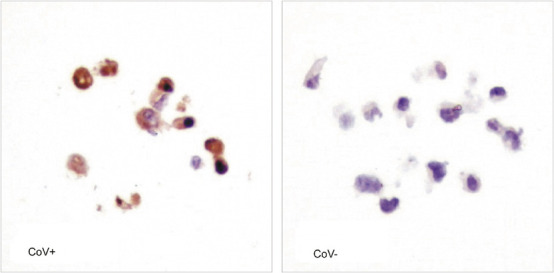
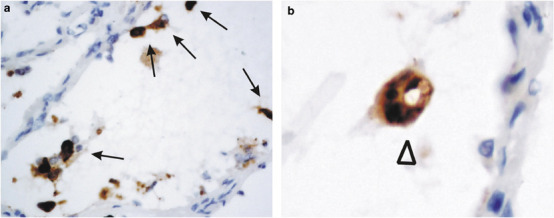
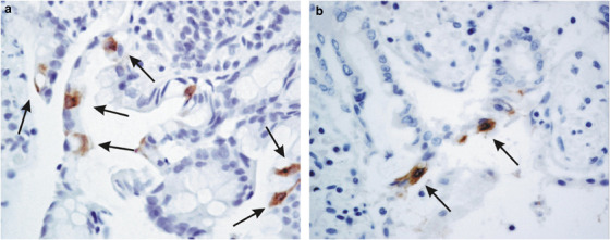
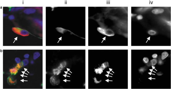
Severe acute respiratory syndrome (SARS) is a newly emerging infectious disease that haunted the world from November 2002 to July 2003. Little is known about the biology and pathophysiology of the novel coronavirus that causes SARS. The tissue and cellular distributions of coronaviral hypothetical and structural proteins in SARS were investigated. Antibodies against the hypothetical (SARS 3a, 3b, 6, 7a and 9b) and structural proteins (envelope, membrane, nucleocapsid and spike) of the coronavirus were generated from predicted antigenic epitopes of each protein. The presence of these proteins were first verified in coronavirus-infected Vero E6 tissue culture model. Immunohistochemical studies on different human tissues, including a cohort of nine autopsies, two liver biopsies and intestinal biopsies of SARS patients, further confirmed the existence of coronaviral hypothetical and structural proteins in the cytoplasm of pneumocytes and small intestinal surface enterocytes in SARS patients. With this vast array of antibodies, no signal was observed in other cell types including those organs in which reverse transcriptase-polymerase chain reactions were reported to be positive. Structural proteins and the functionally undefined hypothetical proteins were expressed in coronavirus-infected cells with distinct expression pattern in different organs in SARS patients. These antipeptide antibodies can be useful for the diagnosis of SARS at the tissue level.
Modern Pathology (2005) 18, 1432-1439. doi:10.1038/modpathol.3800439; published online 13 May 2005.
Figures
References
-
- WHO. Severe Acute Respiratory Syndrome (SARS) last accessed 2004, August 10.http://www.who.int/csr/sars/en/ 2004.
-
- Zhan J, Chen W, Li C, et al. Digestive system manifestations in patients with severe acute respiratory syndrome. Chin Med J (England) 2003;116:1265–1266. - PubMed
Publication types
MeSH terms
Substances
LinkOut - more resources
Full Text Sources
Miscellaneous
