. 2006 Jan;206(1):103-11.
doi: 10.1002/jcp.20433.

EEDA: a protein associated with an early stage of stratified epithelial differentiation

Affiliations

EEDA: a protein associated with an early stage of stratified epithelial differentiation

Lijie Sun et al. J Cell Physiol. 2006 Jan.

Abstract

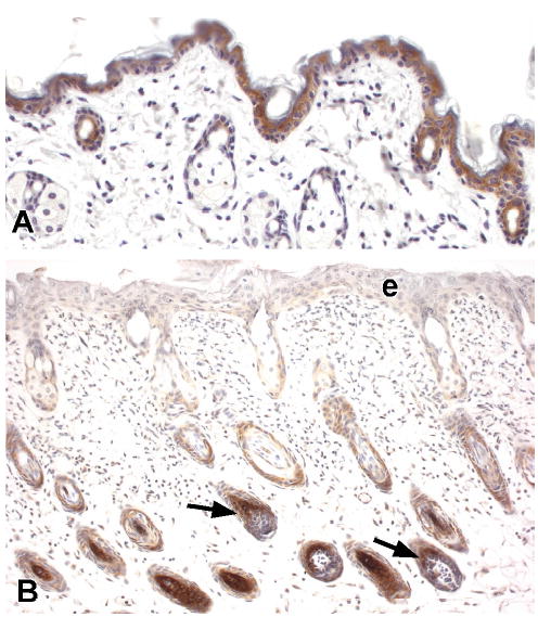
Using suppressive subtractive hybridization, we have identified a novel gene, which we named early epithelial differentiation associated (EEDA), which is uniquely associated with an early stage of stratified epithelial differentiation. In epidermis, esophageal epithelium, and tongue epithelium, EEDA mRNA, and antigen was abundant in suprabasal cells, but was barely detectable in more differentiated cells. Consistent with the limbal location of corneal epithelial stem cells, EEDA was expressed in basal corneal epithelial cells that are out of the stem cell compartment, as well as the suprabasal corneal epithelial cells. The strongest EEDA expression occurred in suprabasal precortical cells of mouse, bovine, and human anagen follicles. Developmental studies showed that the appearance of EEDA in embryonic mouse epidermis (E 15.5) coincided with morphological keratinization. Interestingly, EEDA expression is turned off when epithelia were perturbed by wounding and by cultivation under both low and high Ca2+ conditions. Our results indicate that EEDA is involved in the early stages of normal epithelial differentiation, and that EEDA is important for the "normal" differentiation pathway in a wide range of stratified epithelia.


Figures

Figure 1
Figure 1
The partial nucleotide and deduced amino acid sequences of the cDNA encoding bovine EEDA, an early epithelial differentiation-associated protein. The stop codon is marked with an asterisk.
Figure 2
Figure 2
Alignment of EEDA sequences. Numbers indicate the amino acid positions among the b: bovine; m: mouse; h: human EEDAs. Asterisks denote the following motifs: PKC: protein kinase C phosphorylation site; CKII: casein kinase II phosphorylation site; and N-Gly: N glycosylation site.
Figure 3
Figure 3
Comparison of mouse EEDA with human death-associated protein-1 (DAP-1). Note the high degree of homology between mouse EEDA and human DAP-1.
Figure 4
Figure 4. Northern blot (A) analysis of EEDA mRNAs from various bovine tissues. (Upper panel) m RNAs from various bovine tissues were hybridized with a cDNA fragment of EEDA. An intense signal is observed in the corneal epithelium and a weak signal is seen in the stomach. No signal is seen in the bladder, brain, heart, or liver. (Lower panel) The probe on the same blot was stripped off and rehybridized with a glyceraldehydes-3-phosphate dehydrogenase (GAPDH) cDNA probe as a control
Western blot (B) analysis of a human corneal epithelial extract. Human corneal epithelium was extracted with Tris/Triton-X buffer, resolved on SDS-PAGE, transferred to nitrocellulose membranes, immunoblotted with rabbit polyclonal anti-EEDA antibody and detected with a ECL. A single band is obtained at 12 kD, which is consistent with the molecular weight determined from the Northern blots and confirms the presence of EEDA in human corneal epithelium.
Figure 5
Figure 5. EEDA is expressed in Corneal but not Limbal Epithelium
EEDA mRNA (A, B) and antigen (C, D) were localized in mouse peripheral (A, C) and central (B, D) corneal epithelia. EEDA mRNA and antigen are observed in the basal and wing cells of mouse corneal (co) epithelium, but not in the superficial cells (arrows). Little, if any EEDA can be detected in the limbal (lim) epithelium (A, C); however, in the peripheral corneal (pc) epithelium immediately adjacent to the limbal epithelium EEDA expression is distinct.
Figure 6
Figure 6. EEDA is Expressed Immediately above the Proliferative Zones of the Hair Follicle, Nail Matrix and Dorsal Tongue Epithelium
EEDA mRNA (A, B, C) and antigen (D, E) are localized in bovine (A), human (B) and mouse (C, D) hair follicles and in mouse (E) nails. Within the follicular epithelium (AD), EEDA is detected in the cells immediately above the line of Auber (dashed line), and is of significance in that the bulk of the mitotic activity that gives rise to the hair shaft and inner root sheath occurs in the matrix keratinocytes (m) below this level (Lavker et al., 2003). Within the nail (E) EEDA is seen in the intermediate cells of the nail matrix (nm) but not in the cells of the nail plate (np). Dp, dermal papilla. EEDA antigen (F) is present in the intermediate cells of the posterior (p) portion of the dorsal tongue epithelium. Note lack of EEDA expression in the anterior (a) portion of the epithelium.
Figure 7
Figure 7. EEDA is Expressed Immediately above the Proliferative Zones of Epidermis and Footpad Epithelium
Mouse epidermis (A, B) and footpad epithelium (D, E) were processed for in situ hybridization with 35S-labeled sense (A, D) and antisense (B, E) probes to EEDA. A strong signal for EEDA mRNA is observed in the suprabasal cells immediately above the basal (b) layer in epidermis (B) and footpad epithelium (E). Little if any signal for EEDA mRNA is seen in the granular (g) cells. Inset (E) is a higher magnification of the footpad epithelium showing EEDA mRNA associated mainly with the intermediate cell layers. Mouse epidermis (C) and footpad epithelium (F, G) were processed for immunohistochemical staining using polyclonal rabbit anti-mouse EEDA IgG, biotinylated secondary IgG, the avidin-biotin-peroxidase complex, and the substrate 3,3’-diaminobenzidine. Control experiments were performed using normal rabbit IgG (F). Expression pattern for EEDA antigen is similar to that observed for EEDA mRNA.
Figure 8
Figure 8. EEDA is expressed during the Later Stages of Epidermal Development
EEDA antigen is faintly detected in the two cell epithelium that comprised the E 14.5day (A) epidermis. At embryonic day 15.5 (B) EEDA is easily detected in the cells immediately above the basal (b) layer, but is absent in basal cells and follicular buds (fb). By embryonic day 17.5 (C) EEDA expression is observed above the basal (b) layer and can not be detected in the prominent granular (g) layer. Within the follicular epithelium, EEDA can be observed in an occasional cell (arrow) located at the apex of the matrix (m). EEDA expression pattern in the embryonic 18.5 day skin (D) is similar to that seen in the adult, with strong staining in the suprabasal cells and little if any staining the granular (g) or horny (h) cells. Only those cells (arrow) at the apex of the follicular matrix (m) express EEDA at this time. Dp, dermal papilla
Figure 9
Figure 9. EEDA Expression Becomes Compartmentalized During Neonatal Corneal Epithelial Development
At post-natal day 1(A) EEDA is observed in the single cell limbal (l) peripheral (pc) and central (cc) corneal epithelia. At post-natal day 5 (B, C) EEDA expression is already compartmentalized as evidenced by its absence in the limbal (l) epithelium, faint presence in the peripheral (pc) corneal epithelium and distinct expression in the central (cc ) corneal epithelium. At 9 days of age (D, E) EEDA is expressed in the basal and wing cells, but not in the superficial cells of the corneal epithelium (cc). EEDA expression is also easily observed in the peripheral corneal (pc) epithelium, but not in the limbal (l) epithelium. This pattern of EEDA expression is maintained in the adult (F, G).
Figure 10
Figure 10. EEDA is not expressed in Cultured Keratinocytes
Western blot analysis of epidermal (epi) extracts and extracts of epidermal and corneal keratinocytes cultured under low (0.3μM) and high (1.2 μM) Ca2+ conditions. All lanes were loaded with 20μg total cellular protein. Epidermal EEDA is detected as a single 12kD band, whereas EEDA can not be detected in any of the keratinocyte extracts either before (0 hrs) or after (24, 48, 72 hrs) the addition of Ca2+.
Figure 11
Figure 11. Phorbol Ester Reduces EEDA Expression in the Epidermis
A marked reduction in EEDA expression is observed after 1 (B) day of topical application of 0.01% TPA in petrolatum to the backs of mice compared to topical application of petrolatum (A). Interestingly EEDA expression within the precortical follicular epithelial cells (arrows) is not affected by topical TPA treatment.
Figure 12
Figure 12. Wounding Reduces EEDA Expression in the Epidermis
Penetrating wounds were created on the back skin of adult (7 wk) C57Bl/6 mice. These mice were sacrificed 1 (A, B, C), 3 (D, E, F), 5 (G, H, I) and 7 (J, K, L) days after wounding, and the skin was processed for immunohistochemistry. Immediately following wounding, EEDA expression is markedly reduced in the newly formed epithelium (leading edge; C) and epidermis proximal to the leading edge (near edge; B). As reepithelialization progresses, EEDA expression is initially observed in a patchy pattern, appearing first in the more superficial cells (arrows; E). By 7 days post-wounding EEDA expression in the newly re-epithelialized tissue (L) and the adjacent epidermis (K) appears similar to the unwounded epidermis (J).

Similar articles

Cited by

References

    1. Asselineau D, Bernard BA, Bailly C, Darmon M, Prunieras M. Human epidermis reconstructed by culture: is it "normal"? J Invest Dermatol. 1986;86(2):181–186. - PubMed
    1. Cabodi S, Calautti E, Talora C, Kuroki T, Stein PL, Dotto GP. A PKC-eta/Fyn-dependent pathway leading to keratinocyte growth arrest and differentiation. Mol Cell. 2000;6(5):1121–1129. - PubMed
    1. Chaloin-Dufau C, Sun T-T, Dhouailly D. Appearance of the keratin pair K3/K12 during embryonic and adult corneal epithelial differentiation in the chick and in the rabbit. Cell Diff Dev. 1990;32:97–108. - PubMed
    1. Chen WY, Mui MM, Kao WW, Liu CY, Tseng SC. Conjunctival epithelial cells do not transdifferentiate in organotypic cultures: expression of K12 keratin is restricted to corneal epithelium. Current Eye Research. 1994;13(10):765–778. - PubMed
    1. Cotsarelis G, Cheng SZ, Dong G, Sun TT, Lavker RM. Existence of slow-cycling limbal epithelial basal cells that can be preferentially stimulated to proliferate: implications on epithelial stem cells. Cell. 1989;57(2):201–209. - PubMed

Publication types

MeSH terms

Substances