. 2005 Jun 7;102(23):8192-7.
doi: 10.1073/pnas.0502193102. Epub 2005 May 27.

Heptameric (L12)6/L10 rather than canonical pentameric complexes are found by tandem MS of intact ribosomes from thermophilic bacteria

Affiliations

Heptameric (L12)6/L10 rather than canonical pentameric complexes are found by tandem MS of intact ribosomes from thermophilic bacteria

Leopold L Ilag et al. Proc Natl Acad Sci U S A. .

Abstract

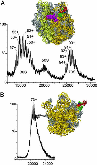
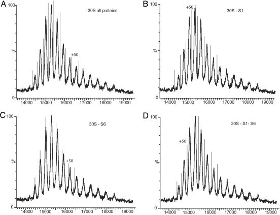
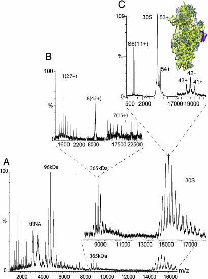
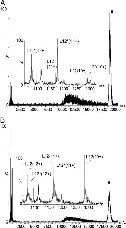
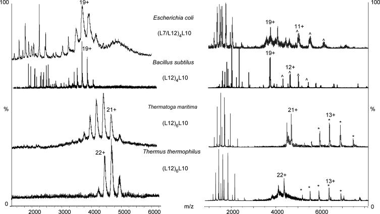
Ribosomes are universal translators of the genetic code into protein and represent macromolecular structures that are asymmetric, often heterogeneous, and contain dynamic regions. These properties pose considerable challenges for modern-day structural biology. Despite these obstacles, high-resolution x-ray structures of the 30S and 50S subunits have revealed the RNA architecture and its interactions with proteins for ribosomes from Thermus thermophilus, Deinococcus radiodurans, and Haloarcula marismortui. Some regions, however, remain inaccessible to these high-resolution approaches because of their high conformational dynamics and potential heterogeneity, specifically the so-called L7/L12 stalk complex. This region plays a vital role in protein synthesis by interacting with GTPase factors in translation. Here, we apply tandem MS, an approach widely applied to peptide sequencing for proteomic applications but not previously applied to MDa complexes. Isolation and activation of ions assigned to intact 30S and 50S subunits releases proteins S6 and L12, respectively. Importantly, this process reveals, exclusively while attached to ribosomes, a phosphorylation of L12, the protein located in multiple copies at the tip of the stalk complex. Moreover, through tandem MS we discovered a stoichiometry for the stalk protuberance on Thermus thermophilus and other thermophiles and contrast this assembly with the analogous one on ribosomes from mesophiles. Together with evidence for a potential interaction with the degradosome, these results show that important findings on ribosome structure, interactions, and modifications can be discovered by tandem MS, even on well studied ribosomes from Thermus thermophilus.


Figures

Fig. 1.
Fig. 1.
Mass spectra of 70S and 50S subunits of ribosomes from Thermus thermophilus. (A) The MS of the 70S was recorded with a 100-V offset on the collision cell and reveals well resolved charge states for the 30S and 70S subunits. The 50S subunit was separated chromatographically, and a spectrum of the isolated subunit B was recorded. The structures of the 70S and 50S particles were produced from the Protein Data Bank coordinates 1GIX and 1GIY, respectively by using the program ribbons (34). The rRNA is shown in gold and yellow for the 50S and 30S subunits, respectively with proteins shown in gray with the exception of those released in mass spectra (see Fig. 2), L12 (red/green) and tRNA pink, red, and green in the A, P, and E sites, respectively.
Fig. 2.
Fig. 2.
Simulation of the isotopes of the 30S subunit. Simulation of the charge states corresponding to various isotopic formulae calculated from the RNA and protein compositions of the 30S subunit is shown. The theoretical charge states are represented as single lines superimposed on the mass spectrum to determine coincidence with the onset of the mass spectral peaks. Simulation of the isotopes was performed with all proteins included (A), in the absence of S1 (B), in the absence of S6 (C), and without both S1 and S6 (D).
Fig. 3.
Fig. 3.
Acceleration of ribosomes from Thermus thermophilus releases individual components and complexes. (A) Mass spectrum of ribosomes from Thermus thermophilus were subjected to an acceleration protocol in which all ions were accelerated at 200 V through the collision cell of the mass spectrometer. This process releases additional complexes and individual proteins and provides enhanced resolution for the 30S subunit. (B) The 365-kDa complex is subjected to tandem MS, the resulting heptamer and monomeric species confirming that the complex is octameric. (C) Tandem MS of the 30S subunit releases a single protein, S6, shown in purple in the Protein Data Bank structure 1GIX (1).
Fig. 4.
Fig. 4.
Tandem MS of the 50S subunit and 96-kDa complex releases L12 and a phosphorylated form of L12. (A) Tandem MS of the 50S subunit releases L12 and a modified form, denoted L12*. (B) After treatment with a phosphatase in the presence of a kinase inhibitor. Peaks assigned to unmodified L12 are more intense than the modified form after phosphatase treatment. Moreover the ratio of proteins released from the 50S parent ion is significantly higher than for the untreated sample.
Fig. 5.
Fig. 5.
Mass spectra of stalk complexes released from different bacterial ribosomes. (Left) Mass spectra of 70S ribosomes from Thermus thermophilus, B. subtilis, and E. coli and 50S subunits from Thermotoga maritima give rise to broad peaks assigned to the stalk complex in each case. For the thermophiles and mesophiles the mass measured is consistent with heptameric and pentameric stoichiometries, respectively. (Right) Tandem mass spectra confirm the stoichiometry of these complexes, giving rise to both L12 and L10 and stripped complexes at high m/z. The charge state that was isolated for tandem MS is labeled with its charge state and the series labeled by * is assigned to a hexameric complex from which L12 has dissociated and ∧ denotes the analogous tetrameric complex.
Fig. 6.
Fig. 6.
Sequence alignment of L10 from Thermotoga maritima, Thermus thermophilus, E. coli, and B. subtilus. Sequence alignment of L10 from Thermotoga maritima, Thermus thermophilus, E. coli, and B. subtilus was carried out with the program clustal w (35). The results reveal an extra eight residues in the C-terminal region of the proteins from the thermophiles compared with E. coli and B. subtilus.

Similar articles

Cited by

References

    1. Wimberly, B. T., Brodersen, D. E., Clemons, W. M., Jr., Morgan-Warren, R. J., Carter, A. P., Vonrhein, C., Hartsch, T. & Ramakrishnan, V. (2000) Nature 407, 327-339. - PubMed
    1. Schluenzen, F., Tocilj, A., Zarivach, R., Harms, J., Gluehmann, M., Janell, D., Bashan, A., Bartels, H., Agmon, I., Franceschi, F. & Yonath, A. (2000) Cell 102, 615-623. - PubMed
    1. Harms, J., Schluenzen, F., Zarivach, R., Bashan, A., Gat, S., Agmon, I., Bartels, H., Franceschi, F. & Yonath, A. (2001) Cell 107, 679-688. - PubMed
    1. Ban, N., Nissen, P., Hansen, J., Moore, P. B. & Steitz, T. A. (2000) Science 289, 905-920. - PubMed
    1. Gudkov, A. T., Tumanova, L. G., Venyaminov, S. Y. & Khechinashvilli, N. N. (1978) FEBS Lett. 93, 215-218. - PubMed

Publication types

MeSH terms

LinkOut - more resources