. 2005 Jun;138(2):819-26.
doi: 10.1104/pp.105.061689. Epub 2005 May 27.

DRP1A is responsible for vascular continuity synergistically working with VAN3 in Arabidopsis

Affiliations

DRP1A is responsible for vascular continuity synergistically working with VAN3 in Arabidopsis

Shinichiro Sawa et al. Plant Physiol. 2005 Jun.

Abstract

In most dicotyledonous plants, vascular tissues in the leaf have a reticulate venation pattern. We have isolated and characterized an Arabidopsis (Arabidopsis thaliana) mutant defective in the vascular network defective mutant, van3. van3 mutants show a discontinuous vascular pattern, and VAN3 is known to encode an ADP-ribosylation-factor-GTPase-activating protein that regulates membrane trafficking in the trans-Golgi network. To elucidate the molecular nature controlling the vein patterning process through membrane trafficking, we searched VAN3-interacting proteins using a yeast (Saccharomyces cerevisiae) two hybrid system. As a result, we isolated the plant Dynamin-Related Protein 1A (DRP1A) as a VAN3 interacting protein. The spatial and temporal expression patterns of DRP1AGUS and VAN3GUS were very similar. The subcellular localization of VAN3 completely overlapped to that of DRP1A. drp1a showed a disconnected vascular network, and the drp1a mutation enhanced the phenotype of vascular discontinuity of the van3 mutant in the drp1a van3 double mutant. Furthermore, the drp1 mutation enhanced the discontinuous vascular pattern of the gnom mutant, which had the same effect as that of the van3 mutation. These results indicate that DRP1 modulates the VAN3 function in vesicle budding from the trans-Golgi network, which regulates vascular formation in Arabidopsis.


Figures

Figure 1.
Figure 1.
VAN3 interaction with DRP1A. A, Yeast two hybrid analysis. The Y190 yeast strain was transformed with pAS2-VAN3 and pACT2-DRP1A. The yeast harboring pAS2-DRP1A and pACT2-VAN3 were grown on agar plates without His. The Y190 yeast strain was transformed with pAS2-VAN3 and the pACT2 vector or pAS2-DRP1A and the pACT2 vector are shown as negative controls. B, Protein-protein interaction assay with GST pull down. VAN3-GAL4 DNA binding domain pull down with purified GST and GST-DRP1A used as bait. The VAN3 protein was detected with a western-blot analysis. C, Time course of DRP1A GTPase activity. Recombinant DRP1A protein was incubated with 5 μm [α -32P] GTP. GTP hydrolysis was observed (*) in the left section. GTP hydrolysis was quantitatively analyzed with a BIO-imaging analyzer in the right section.
Figure 2.
Figure 2.
Genetic interaction of the drp1a mutant. A, Wild type. B, About 10% of drp1a mutants show a disconnected vasculature. Arrows show the vascular disconnection. C, van3 mutants show a discontinuous vasculature. D, drp1a van3 double mutants showed a severe defect in vascular development. Arrows show the restricted maturation of the tracheary element. E, gnom mutants show a concentrated vasculature. F, gnom van3 double mutants show a highly fragmented vasculature. G, gnom drp1a double mutants show a fragmented vasculature similar to the gnom van3 double mutant. Bars = 500 μm.
Figure 3.
Figure 3.
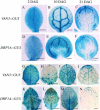
Temporal and spatial expression pattern of DRP1A and VAN3 during leaf development. Histochemical localization of VAN3GUS (A–C, G–J) and the DRP1AGUS (D–F, K–N) reporter gene expression in the cotyledon (A–F) and in rosette leaf (G–N). Samples were fixed and incubated in a reaction buffer including 2 mg/mL of X-Gluc for 10 min. Bars = 500 μm (A–F), 1 mm (G–N).
Figure 4.
Figure 4.
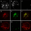
Subcellular localization of DRP1A and VAN3. 35S-promoter-driven RFP-tagged DRP1A. Arrows show punctate structures. A, GFP-tagged MAP4 as a microtubule marker (B) and VENUS-tagged VAN3 (C) were introduced into protoplasts of Arabidopsis suspension-cultured cells. RFP-DRP1A and GFP-MAP4 (D–F), and RFP-DRP1A and VAN3-Venus (G–I) were cointroduced. Localization of RFP-DRP1A (D), MAP4-GFP (E), and merged image (F) are shown. The location of RFP-DRP1A (G), VAN3-VENUS (H), and merged image of RFP-DRP1A and VAN3-VENUS (I) are shown. Fluorescence was observed by confocal laser microscopy (LSM510 META, Carl Zeiss).

References

    1. Aloni R (2001) Foliar and axial aspects of vascular differentiation: hypotheses and evidence. J Plant Growth Regul 20: 22–34
    1. Bednarek SY, Falbel TG (2002) Membrane trafficking during plant cytokinesis. Traffic 3: 621–629 - PubMed
    1. Berleth T, Mattsson J, Hardtke CS (2000) Vascular continuity and auxin signals. Trends Plant Sci 5: 387–393 - PubMed
    1. Busch M, Mayer U, Jürgens G (1996) Molecular analysis of the Arabidopsis pattern formation gene GNOM: gene structure and intragenic complementation. Mol Gen Genet 250: 681–691 - PubMed
    1. Carland FM, Berg BL, FitzGerald JN, Jinamornphongs S, Nelson T, Keith B (1999) Genetic regulation of vascular tissue patterning in Arabidopsis. Plant Cell 11: 2123–2137 - PMC - PubMed

Publication types

MeSH terms